Download
1 / 78
790 likes | 1.39k Views
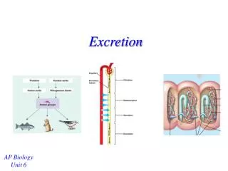
Osmoregulationand Excretion. Osmoregulation : Regulation of solute concentrations and water balance by a cell or organism. Osmoregulation balances the uptake and loss of water and solutes. . If cells uptake too much water they will burst

E N D
Osmoregulationand Excretion
Osmoregulation: Regulation of solute concentrations and water balance by a cell or organism. • Osmoregulation balances the uptake and loss of water and solutes.
If cells uptake too much water they will burst • If cells uptake too little water, or lose too much water they will shrivel and die. • Osmoregulation is impacted by water intake/loss and also discharge of metabolic wastes.
Osmosis: movement of water across a selectively permeable membrane when two solutions separated by the membrane differ in osmotic pressure. Initial flaccid cell 0.4 M sucrose solution Distilled water
Osmolarity(osmotic pressure): Total solute concentration expressed as molarity (moles of solute per liter of solution). • Unit of osmolarity: milliOsmoles per liter (mOsm/L); equivalent to solute concentration of 10-3M. • Human blood: 300 mOsm/L • Seawater: 1000 mOsm/L
Isotonic: two solutions on either side of the membrane have same osmolarity; no net movement of water between two sides. • Hyperosmotic/ hypertonic: the solution with greater solute concentration (net movement of water towards this side) • Hypoosmotic/ hypotonic: the more dilute solution (net movement of water away from this side) • An animal has to have a way of maintaining water balance with its environment.
Osmoconformer: • Isoosmotic with the surroundings • Internal osmolarity same as the environment • No net gain or loss of water • All marine
Osmoregulator: • Maintains its osmolarity independent of the environment. • Because of the regulatory capacity can live in freshwater, dry land, also marine habitats • In hypoosmotic environment they discharge excess water • In hyperosmotic environment they take up and retain more water.
Based on what you know so far about thermoregulation can you make any guesses about osmoregulation in these two groups? • Balancing act between control and cost.
Stenohaline: Cannot tolerate substantial changes in external osmolarity • Euryhaline: Can tolerate large fluctuations in osmolarity (organisms in intertidal zones, fishes that migrate between sea water and fresh water) • Osmoregulators and osmoconformers can be found in both these groups.
Marine invertebrates: osmoconformes, need to transport some solutes to maintain homeostatis.
Marine bony fishes: • lose water by osmosis • drink large amounts of water • actively transport out Cl- (chloride cells) • passively transport out Na+ • concentrated urine gets rid of other salts like Ca++, Na+, Mg++
Gain of water and salt ions from food and by drinking seawater Osmotic water loss through gills and other parts of body surface Excretion of salt ions and small amounts of water in scanty urine from kidneys Excretion of salt ions from gills Osmoregulation in a saltwater fish
Marine cartilaginous fishes: • Shark tissue contains a high concentration of urea • To prevent urea from damaging other organic molecules in the tissues they have trimethyl amine oxide (TMAO) • Because of high solute concentration in tissue water enters the cells (sharks don’t drink) • Produce concentrated urine.
Fresh water organisms: (opposite problem of marine organisms) • Internal osmolarity is higher than surroundings; problem of gaining water. • Fishes don’t drink water , large volumes of urine • Salt intake through food • Chloride cells in gills actively transport in Cl- • Na+ follows
Osmotic water gain through gills and other parts of body surface Uptake of water and some ions in food Uptake of salt ions by gills Excretion of large amounts of water in dilute urine from kidneys Osmoregulation in a freshwater fish
Euryhaline organisms like salmon: • In sea they drink sea water and discharge salt through their gills • In freshwater they stop drinking and produce large volumes of dilute urine, gills take up salt Life Cycle of Atlantic Salmon http://www.nefsc.noaa.gov/sos/spsyn/af/salmon/images/fig41_2.gif
Anhydrobiosis: dormant state when habitat dries up. 85% to 2% water in water bears. Cell membrane adaptations are poorly understood.
100 µm 100 µm Dehydrated tardigrade Hydrated tardigrade
Land animals: • Adaptations of body surface (thick cuticle) and behavior (nocturnal) help reduce water loss. • Some desert animals can metabolically generate water (kangaroo rats)
Water balance in a kangaroo rat (2 mL/day) Water balance in a human (2,500 mL/day) Ingested in food (750 mL) Ingested in food (0.2 mL) Ingested in liquid (1,500 mL) Water gain Derived from metabolism (1.8 mL) Derived from metabolism (250 mL) Feces (100 mL) Feces (0.09 mL) Urine (1,500 mL) Urine (0.45 mL) Water loss Evaporation (900 mL) Evaporation (1.46 mL)
Transport epithelia: Animals that live on sea water can also eliminate salt through specialized epithelial cells that can regulate the salt concentration.
Nasal salt gland Nostril with salt secretions
Nitrogenous wastes: • As a result of metabolism proteins and amino acids produce ammonia (NH3). • Ammonium ion (NH4+) is highly toxic • Animals either get rid of ammonia promptly or expend energy and convert it to less toxic forms.
Forms of nitrogenous wastes: • ammonia • urea • uric acid (differ in cost to convert and toxicity)
Ammonia: • Most fishes, animals that produce shell-less eggs. • Excrete bulk of ammonia through gills, minor amounts through kidneys. • Very toxic, has to be transported in very dilute solutions
Urea: • Mammals, adult amphibians, some marine , bony fishes, sharks, turtles. • Advantage: Lower toxicity. Can go through circulatory system, stored. Does not have to be so dilute, so less water loss during excretion. • Disadvantage: High energy cost. • Animals can switch mode of excretion at different stages of their life cycle. Tadpoles (ammonia), adult amphibians (urea).
Uric acid: • reptiles, birds, land insects, animals that produce shelled eggs. • Advantage: less toxic than urea, needs less water to be excreted (semisolid paste). • Disadvantage: more expensive to produce than urea. • Humans produce small amounts of uric acid. Gout: condition caused by inability to eliminate uric acid.
Nucleic acids Proteins Amino acids Nitrogenous bases —NH2 Amino groups Most aquatic animals, including most bony fishes Mammals, most amphibians, sharks, some bony fishes Many reptiles (including birds), insects, land snails Ammonia Urea Uric acid
Amount of nitrogenous waste: linked to energy budget (higher in endotherms than ectotherms).
LE 44-9 Capillary Filtration Excretory tubule Filtrate • Steps in urine formation: • Filtration • Reabsorption • Secretion Reabsorption Secretion Urine Excretion
Filtration: • Cells, large molecules (proteins) stay in the body fluid • Small molecules, (salts, sugars, amino acids, nitrogenous wastes) and water pass through and form filtrate
Reabsorption : • Selective process. Recovery of useful molecules • Active transport – reabsorption of certain salts, vitamins, hormones, amino acids • Wastes, nonessential molecules are left behind
Secretion: • Selective pumping of various solutes to adjust osmotic movement of water into and out of the filtrate • Final step – removal of this filtrate from the body – release of urine
Diverse excretory systems: • Excretory system plays a very important role in water balance and homeostasis. • Systems show a lot of variation in different groups • Basic structure – network of tubules that provide a large surface area
Nucleus of cap cell Protonephridia - excrete low concentration of solute; flatworms, some rotifers, some annelids and molluscs Cilia Interstitial fluid filters through membrane where cap cell and tubule cell interdigitate (interlock) Tubule cell Flame bulb Protonephridia (tubules) Tubule Nephridiopore in body wall
Metanephridia - found in most annelids (e.g. earthworms); excretory organs open internally to the coelom Coelom Capillary network Bladder Collecting tubule Nephridio- pore Nephrostome Metanephridium
Malpighian tubules – • extend from hemolymph to digestive tract; • cells secrete nitrogenous wastes and other solutes into hemolymph, • these molecules and water move into malpighian tubules, • excess water is reabsorbed in the rectum; • other essential solutes are reabsorbed; • lets the animal conserve water; • found in insects, capability to conserve water helps in the success of this group
Digestive tract Rectum Hindgut Intestine Midgut (stomach) Malpighian tubules Anus Feces and urine Salt, water, and nitrogenous wastes Malpighian tubule Rectum Reabsorption of H2O, ions, and valuable organic molecules HEMOLYMPH
Kidneys: vertebrates and some other chordates, same basic plan as other systems – but highly organized and complex, closely associated with a network of capillaries.
Posterior vena cava Renal artery and vein Kidney Renal medulla Aorta Renal cortex • Structure of mammalian excretory system Ureter Renal pelvis Urinary bladder Urethra Ureter Excretory organs and major associated blood vessels Section of kidney from a rat Kidney structure Juxta- medullary nephron Cortical nephron Afferent arteriole from renal artery Glomerulus Bowman’s capsule Proximal tubule Peritubular capillaries Renal cortex Collecting duct SEM 20 µm Efferent arteriole from glomerulus Renal medulla Distal tubule To renal pelvis Collecting duct Branch of renal vein Descending limb Loop of Henle Nephron Ascending limb Vasa recta Filtrate and blood flow
Kidney Renal medulla Renal cortex Renal pelvis Ureter Section of kidney from a rat Kidney structure
Juxta- medullary nephron Cortical nephron Renal cortex Collecting duct Renal medulla To renal pelvis Nephron
Afferent arteriole from renal artery Glomerulus Bowman’s capsule Proximal tubule Peritubular capillaries SEM 20 µm Efferent arteriole from glomerulus Distal tubule Collecting duct Branch of renal vein Descending limb Loop of Henle Ascending limb Vasa recta Filtrate and blood flow
Detailed look at processing of blood in nephron: • Filtration • Afferent arteriole has a bigger diameter than efferent arteriole, thus pressure builds in the glomerulus • During filtration, blood in glomerulus is forced into Bowman’s capsule (cup shaped swelling at the blind end of the tubule) • Filtration is nonselective, only based on size, caused by the high blood pressure in the capillaries in the Bowman’s capsule.