Download
1 / 11
120 likes | 433 Views
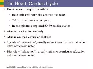
Heart sounds and intra-cardiac pressures. Heart sounds:. S1: Due to closure of mitral and tricuspid valves. Softer: Cardiac failure, pericardial effusion, valvular incompetence Louder: Tachycardia, shortened PR-interval, mitral stenosis with still pliable valve.

E N D
Heart sounds: • S1: Due to closure of mitral and tricuspid valves. • Softer: Cardiac failure, pericardial effusion, valvular incompetence • Louder: Tachycardia, shortened PR-interval, mitral stenosis with still pliable valve
S2: Closure of aortic and pulmonary valves. • Louder: Systemic and/or pulmonary HT • Softer: Pericardial effusion, hypotension, valvular incompetence
Splitting of S1 and S2: • Normally: Deep inspiration, RV-filling, split S1: Tricuspid valve closes after mitral valve. Physiologicl splitting of S1. • ASD: Fixed splitting of S1 • Electrical disturbances: RBBB, LBBB
Splitting of S2: • Any condition(s) impairing emptying of LV or RV. • Valvular stenosis, pulmonary HT etc
S3: • Caused by rapid ventricular filling • Can be normal in the young • Pathology: • Valvular incompetence
S4: • Caused by stiff ventricle • During atrial contraction • AF: S4 ? or not
Intra-cardiac pressures: • Intra-atrial pressure: RA: a,c,v waves on flebogram: From negative to +8 mmHg • LA: Also a,c,v waves: Maximum +12 mmHg: Measured by catheter: Swan-Ganz • Pulmonary capillary wedge pressure:
Clinical application of wedge pressure: • Pulmonary edema: Cardiac vs non-cardiac • Increased wedge pressure=cardiac origin: • LV-failure or mitral valve: stenosis or acute incompetence • LV-failure: Systolic vs diastolic
Intra-ventricular pressures: • RV: From 0 to 25 mmHg • LV: From 0 to 125 mmHg
LV pressure-volume curve • Diagram on cardiac cycle • La Place`s law • Poiseulle`s law • Study from textbook: Ganong