Download
1 / 59

E N D
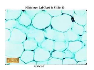
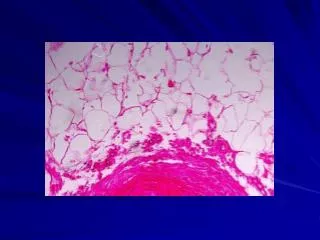
Adipose Tissue This histology slide shows adipose tissue surrounding the ureter. The adipocytes are shaped like a signet ring. The nucleus is pushed to the periphery of the cell. The staining process leaches the fat out of the cells, so these are just the skeleton of the cell. Some smooth muscle from the ureter is visible in this section as well.
Fat cells -- note nucleus and rim of cytoplasm pushed to one side by the accumulation of fat. The lipid itself has been dissolved out in fixation. In the center of the picture, in the space bounded by the four large fat cells, there is a small, round cross-cut of a capillary with a dark, shrunken red blood cell inside.
Dense irregular c.t. (blue) packing around a nerve bundle. The coat immediately surrounding the whole nerve bundle is particularly dense and consists mainly of collagen fibers. In between the individual pale, round nerve fibers is a very fine areolar c.t. packing, with mainly reticular fibers
Dense irregular c. t., with fibers running in all directions. The fibers are mainly collagenous, but keep in mind that some would be elastic and can be seen only if specifically stained. This kind of c.t. is found where firmer packing and binding is needed. The two arrows at top of picture are pointing to elongate, dark, fibroblast nuclei.
A stretched preparation of areolar connective tissue. The pink fibers of different thicknesses are collagenous (or white) fibers. The dark, thin, more tortuous fibers are elastic (or yellow) fibers. Most of the nuclei belong to fibroblasts
Areolar c.t. immediately underlying simple columnar epithelium. This is a very cellular variety of areolar c.t., with a high population of lymphocytes.
Loose irregular connective tissue (also called areolar tissue) as seen underlying and supporting epithelium in an ordinary section. It is rather cellular and supports many small blood vessels which travel through it.
Loose (areolar) connective tissue - (in blue) - surrounding the epithelium of tubules. In areas like this, the finest collagen fibers lying closest to the tubules would be reticular fibers; the only way to distinguish them here from heavier collagen fibers would be to silver them. (The blue here simply stains collagen in general.) REMEMBER: in an area like this, reticular fibers (like all other fibers) are produced by fibroblasts. Only in the primitive reticular tissue of bone marrow, lymph node, and spleen are reticular fibers produced by reticular cells
Reticular tissue (silvered, black). A network of very fine reticular fibers can be seen here, forming the stroma (framework) of a lymph node. These fibers are produced by reticular cells. The pale cells seen in the meshes of the reticular fibers are lymphocytes
7a • Obove
Tendon, cut in cross-section. The pale pink background represents the cut ends of bundles of thick collagen fibers, very closely packed together. The wispy lines you see throughout are the "cracks" between fiber bundles. In the cracks lie fibroblasts which often look triangular or stellate because of being squeezed between the fibers.
Areolar c.t. -- the thin cell running diagonally toward the lower right from the center is a fibroblast
Another fibroblast -- in the curve of the pink collagen fiber. The long, narrow nucleus is characteristic.
Several fibroblasts, lying among collagen fibers. Hardly any cytoplasm is visible.
Fibrocartilage Even though fibrocartilage is a type of cartilage, conceptually it can be thought of as a blend of dense connective tissue and cartilage. The chondrocytes sitting in lacunae are apparent. Bundles of collagen fibers are visible on this histology slides
Hyaline Cartilage These ares histology sides of hyaline cartilage. All the slides demonstrate chondrocytes sitting in lacunae.
Mesenchyme -- embryonic c.t. with multipotential cells. The stellate cells are beginning to form fibers. Sometimes cells are more spindle shaped. Ground substance material is watery and invisible.
Two large macrophages (one on either side of the picture) -- with engulfed particles of blue dye in their cytoplasm. Their nuclei are pink. Compare the irregular sizes of the blue phagocytized particles here with the more even-sized granules of the mast cells in the next two slides. Notice also that the particles in the macrophage are scattered randomly
Mast cells -- deep purple metachromatic stain for granules. Again, granules are spilling out as a result of the preservation techniques. Notice how round and seed-like the granules are and how tightly they are packed in the cell. The cell nuclei are light blue.
Plasma cell -- with somewhat basophilic cytoplasm and an eccentric nucleus with dark blocks of chromatin in it. Note the pale cytoplasmic area to the left of the nucleus; this is the negative Golgi body. Note also the pink collagen fibers scattered irregularly throughout the pale ground substance of the whole field, which is typical of areolar connective tissue
Eosinophils (bright pink granules) -- in areolar connective tissue. Note the bilobed nucleus in the center cell. Pale oval nuclei in the upper left hand corner probably belong to fibroblasts. Small, dark, round nuclei, such as in the lower right quadrant, probably belong to lymphocytes. Macrophages are hard to identify unless their cytoplasm is filled with phagocytized particles.