Download
1 / 10
100 likes | 314 Views
Lab-on-a-chip Devices for Cell Separation and Identification. Feb 2008. A PhD project at Macquarie University under the guidance of Dr. David Inglis and Prof. Ewa Goldys. Microbes, cells and small organisms come in a wide array of shapes and sizes. Yeast. Blood Cells.

E N D
Lab-on-a-chip Devices for Cell Separation and Identification Feb 2008 A PhD project at Macquarie University under the guidance of Dr. David Inglis and Prof. Ewa Goldys.
Microbes, cells and small organisms come in a wide array of shapes and sizes Yeast Blood Cells Photo Credit: H.D.A Lindquist, U.S. EPA cryptosporidium
Microfluidic or Lab-on-a-chip Devices can be used to separate different cell types • Various blood cell separations have been demonstrated: • J. A. Davis, D. W. Inglis, K. M. Morton, D. A. Lawrence, L. R. Huang, S. Y. Chou, J. C. Sturm, and R. H. Austin, Deterministic hydrodynamics: taking blood apart, Proc. Nat. Acad. Sci. (USA), vol. 103, pp. 14779-14784, 2006. • D. W. Inglis, J. A. Davis, T. J. Zieziulewicz, D. A. Lawrence, R. H. Austin, and J. C. Sturm, Determining blood cell size using microfluidic hydrodynamics, J. Immunol. Meth., vol. 329, pp. 151-156, 2008. • D. W. Inglis, K. J. Morton, J. A. Davis, T. J. Zieziulewicz, D. A. Lawrence, R. H. Austin, and J. C. Sturm, Microfluidic device for label-free measurement of platelet activation, Submitted to Lab on a Chip, Jan 08. • But no work yet with microorganisms!
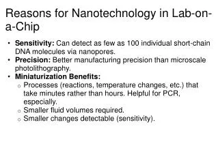
This project will lead to publications which demonstrate the separation of microorganisms on portable inexpensive polymer microfluidic devices. Such devices may have applications in • early detection of human diseases such as sepsis and various parasitic diseases • as well as management of swimming pool and drinking water quality.
What is Micro-Fluidics Fabrication Process: Photolithography • Microfluidics –microfabricated structures for fluid handling • Engineers know how to microfabricate and build integrated systems • mTAS – Micro Total Analysis System • Integrate microfluidic components into a useful device, a “Lab on a Chip” • “development and application of micro- and nanofabricated devices and systems for chemical and biochemical measurements. The technology encompasses extensive areas of application in clinical diagnostics, genomics and proteomics, environmental assays, separation science, cellular analysis or drug discovery.” From Micralyne.com • Interdisciplinary – Physics, Chemistry, Biology Reactive Ion Etching Drill access holes Seal with cover slip Fluid goes in
1.4 mm 1 mm 1.5 mm 1.6 mm 200 mm 1.7 mm 1.8 mm 1.9 mm 2.0 mm 2.1 mm 2.2 mm High Resolution Separation with Size - Position Correlation • Peak width ~10 nm • No known method fractionates with such resolution. Can include chirp to fractionate over a range of sizes by varying gor Huang et al, Science 2004
White blood cells come in different sizes and can be differentiated ~ Cancerous Lymphocytes Healthy Lymphocytes
Human platelets change size in response to chemical and temperature stimulus
Proposed Device • Use the technique described above to create a simple to use high throughput cell concentrator. • Parallel channels for separation and concentration on a glass slide. A second layer distributes fluid to the channels and collects their output. • Structures will be molded PDMS. • Master mold will be lithographically defined SU-8 polymer. 20-micron PDMS pillars