Download
1 / 25
290 likes | 606 Views
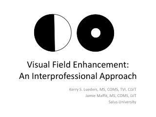
Visual Field Examinations. Week 7 Review. Visual field examinations review. Perimetry - the science of measuring the peripheral vision in order to determine the visual field. (The art of determining boundaries of the visual field.)

E N D
Visual Field Examinations Week 7 Review
Visual field examinations review • Perimetry- the science of measuring the peripheral vision in order to determine the visual field. (The art of determining boundaries of the visual field.) • Visual field- the portion of visual space which is visible to an individual at any given moment. • Isopter-Boundaries of VF areas of equal or greater sensitivity. Isopters are used in Goldmannperimetry. Each isopter is labeled with the size and intensity of the stimulus.
Visual field review • Threshold-A stimulus intensity which has a 50% probability of being seen. This test compares what has the probability of being seen with the possible inaccuracy of the test. Computerized perimetry only. • Asb- intensity value of a stimulus. Used in static perimetry. Smaller asb= greater retinal sensitivity. • dB- intensity value of a stimulus. Larger dB= greater retinal sensitivity.
Visual field examinations review • Scotoma • Visual loss, either blindness (absolute) or decreased sensitivity (relative). scotoma
Visual field examinations review Island of vision An analogy for visual field testing.
Visual field examinations review • 95-100 degrees=temporal • 75 degrees= inferior • 60 degree= nasal • 60 degrees=superior Physiological blind spot Normal visual field boundaries
30-2 and 24-2 threshold- white lights, varying in intensity. Tests 30 or 24 degrees off the macula in all meridians on dilated patient. Macula- white lights, varying in intensity. Tests 5 degrees off the macula in all meridians on dilated patient. Plaquinil- red lights, varying in intensity. Tests 5 degrees of the macula in all meridians on dilated patient. Used for the drug “Plaquinil” which can damage cone photoreceptors.(RA) Bleph- White lights, fixed intensity testing the superior region of the visual field on an undilated patient. There is a lower fixation light. False positive-no light shown, patient responded. (“trigger happy”.) False negative-no response to a previously seen target. Visual field examinations review Computerized VF tests
Computerized visual field testing 30-2 threshold VF Central 10/macular VF
Screening tests • Minimal information done on a computerized VF. • Takes much less time to perform. • C= central region • P= peripheral region • FF= full field • The number after the letter indicates how many targets will be shown to the patient. • The targets are fixed intensity. • It does not print out in grey scale- screening tests will show a circle for seen targets and a black square for unseen targets. Visual field examinations review Computerized VF tests
Visual field examinations review Confrontation visual field Amsler grid
Visual field examinations review Technician moves target, changes target intensity and size, watches fixation, plots chart, investigates a depression or scotoma. Goldmann VF testing can be done with static or kinetic targets. Isopters are used and are color coded or labeled to indicate intensity of target. Goldmann manual VF
Hemianopia- • Binocular- • Monocular- • Homonymous- • Congruent- • Incongruent- • Bi nasal- • Bi temporal- • Altitudinal- • Central cecal- • Nasal step-( bjerrum) • Temporal wedge- Visual field examinations review Types of scotomas
Visual field examinations review Types of scotomas
Monocular retinal zone • Nerve fiber/ optic nerve zone • Binocular chiasmal zone • Post chiasmal zone Visual field examinations review 4 visual pathway zones
Monocular retinal zone • This zone includes the retinal layer, sub retinal layer, rod and cone dystrophy or damage, retinitis pigmentosa, and macular pathology. • Characteristics • All defects will be monocular. • Most pathology will be visible with a scope. • Lesions temporal to fovea present nasal of VF. (inverted field image) • Lesions can cross meridians. • Central scotomas will cause abnormal VA and color vision will be compromised. Visual field examinations review 4 majors visual pathway zones
Nerve fiber/optic nerve zone • This zones includes glaucoma, papilledema, and nerve fiber bundle defects. • Characteristics • All defects will be monocular. • Both eyes can have the same disease causing congruent or incongruent scotomas. • Most defects will be visible with a scope. • Defects will points to the disc since nerve fibers are traveling towards this point. • Glaucomatous scotomas will respect the horizontal meridian. Visual field examinations review 4 major visual pathway zones
Binocular chiasmal zone • This zone includes pathology or damage to the pituitary gland. • Charateristics • Defects will be bilateral, most commonly bi temporal but bi nasal defects can also occur. • NOT visible with a scope, a CT-scan or MRI will be ordered. • Defects here are not an eye problem, this problem is in the brain and effects both eyes because the nasal axons or nerve fibers have crossed to the opposite side at the chiasm. • Defects will respects the vertical meridian and be hemianopic. Visual field examinations review 4 major visual pathway zones
Post chiasm zone • This zone includes the optic tract, lateral geniculate body, optic radiations and the occipital cortex. • Characteristics • Most defects will respect the vertical meridian. • Defects will be homonymous and hemianopic. • This is NOT an eye problem, this is a brain problem and a CT-scan or MRI will be ordered. • Nerve fibers from the nasal VF of one eye and temporal fibers of the other eye will be effected on one side of the brain giving the defect a classification of left or right even though is effects both eyes. The left or right relates to the side of the brain effected. Visual field examinations review 4 major visual pathway zones
Visual field examinations review 4 major visual pathway zones
Thank you! I had a great time teaching you! Good luck on the test and have a great summer! See you in September.