Download
1 / 14
310 likes | 1.16k Views
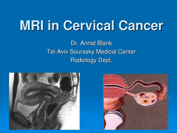
MRI in Cervical Cancer. Dr. Annat Blank Tel-Aviv Sourasky Medical Center Radiology Dept. MR imaging protocol. Pelvic surface array multi channel coil (improved SNR) Fasting or Antispasmodic (optional) FOV 20-24 cm, 3-5 mm slice T2W FSE Axial, sagittal, coronal Axial T1W to renal hilum

E N D
MRI in Cervical Cancer Dr. Annat Blank Tel-Aviv Sourasky Medical Center Radiology Dept.
MR imaging protocol • Pelvic surface array multi channel coil (improved SNR) • Fasting or Antispasmodic (optional) • FOV 20-24 cm, 3-5 mm slice • T2W FSE • Axial, sagittal, coronal • Axial T1W to renal hilum • Lymphadenopathy • Sagittal and axial - Dynamic and delayed C+ Fat suppression • MR spectroscopy, lymph node-specific contrast agents – not rutine.
Cervical Ca - (new) FIGO staging • Stage I - confined to uterus (extension to corpus should be disregarded). • IA – Invasive carcinoma diagnosed only by microscopy • IB - Clinically visible lesion confined to the cervix or microscopic lesion > 5*7mm • IB1 – Clinically visible lesion ≤4.0 cm in greatest dimension. • IB2 – Clinically visible lesion >4.0 cm in greatest dimension • Stage II - invading beyond the uterus but not to the pelvic wall or lower 1/3 of the vagina • IIA - No parametrial involvement. • IIA1 - Clinically visible lesion ≤4.0 cm in greatest dimension. • IIA2 - Clinically visible lesion >4.0 cm in greatest dimension • IIB: - Parametrial involvement, but not onto the pelvic sidewall. • Stage III - cervical cancer invading to the pelvic wall and/or lower 1/3 of the vagina and/or causing a non-functioning kidney • IIIA: No extension onto the pelvic sidewall but involvement of the lower third of the vagina. • IIIB: Extension onto the pelvic sidewall or hydronephrosis or nonfunctioning kidney • Stage IV is carcinoma that has extended beyond the true pelvis or has clinically involved the mucosa of the bladder and/or rectum. • IVA - cervical cancer that invades the bladder or rectum, or extends beyond true pelvis • IVB - distant metastases (distant nodes, lung, liver, bone etc)
“After cervical cancer has been diagnosed, tests are done to find out if cancer cells have spread within the cervix or to other parts of the body”.National Cancer Institute Nov 2011 Which imaging modality? • IVP, Barium enema, rectoscopy cystoscopy,CXR, Ultrasound Lymphangiogram (???) • MRI • PET CT • CT scan (FNA)
Cervical Ca – MR staging accuracy MRICT • Overall 75-96% 50-88% • Parametrial inv. ~90% 30-58% • Lymph nodes 86-88% 85-90% • Stages III-IV 85-95% 90-92%
Cervical Ca – Impact of MR staging • Accurate for pre-op evaluation • Parametrial involvement: sens. 69%, spec. 93% NPV-94-100% • Cost minimizing • Fewer tests • Fewer invasive procedures Radiology 1996; 198: 403
Recurrent Cervical Ca • Pelvic Exenteration may be curative (25-65%) - Need to pick surgical candidates carefully • No sidewall invasion • No extrapelvic mts.
Recurrent Cervical Ca – MRI • High SI on T2W images • Accuracy 80% (DD: inflammatory, edema) • NPV 97% • Dynamic Contrast enhanced • Accuracy 83% • NPV 86% Manfred et al. Radiology 1998 ; 209: 819
Diffusion-weighted MR • Diffusion-weighted magnetic resonance (DWI) imaging is a technique whose contrast derives from the random motion of water molecules within tissues • In solid tumors of high cellularity and extracellular space of tumors there are restrictions to free water movement • Functional not just morphological imaging • Cervical cancers - significantly lower apparent diffusion coefficient (ADC) values compared with normal cervical tissue
CONCLUSION • MRI , although not officially incorporated in the FIGO staging system, has a critical tool in the work-up of cervical carcinoma • Widely accepted as a reliable tool for staging treatment planning and follow-up. • MR can identify prognostic factors.
CONCLUSIONAdvantages of MRI • Tumor size/volume – esp. if tumor is endocervical (MRI>CT) • Stormal and parametrial invasion (MRI>CT) • Nodal Mts. - Periaortic and pelvic LN (PET CT>MRI) • MRI – size criteria , PET CT – sens. for nl sized LN
CONCLUSIONCervical Ca – Imaging guidelines • MRI should be the initial exam for surgical candidates • If MRI is equivocal for bladder or rectum involvement - Endoscopy • PET-CT for distant staging • CT for percutaneous LN biopsy
References Kurts et at. Radiology 1999; 212: 19-27 Yang et al AJR 2000; 175: 759 Nicolet et al RadioGraphics 2000; 20:1539–1549 Okamoto et al RadioGraphics 2003; 23:425–445 Jeong et al RadioGraphics 2003; 23:969–981 Woodward et al Radiographics 2004; 24:225-246 Sala et al AJR 2007; 188:1577-1587 Viswanathan et al Radiographics 2008; 28:289-307 Whittaker et al Radiographics 2009; 29:759-778