Download
1 / 57
570 likes | 758 Views
Patella Fractures & Extensor Mechanism Injuries. OTA Resident’s Course 2005. Lisa K. Cannada, MD Original Authors: Charles G. Haddad, Jr., MD, Lisa K. Cannada, MD, and Robert Cantu, MD; March 2004 New Author: Lisa K. Cannada, MD; Revised January 2006. Anatomy. Largest sesamoid bone

E N D
Patella Fractures & Extensor Mechanism Injuries OTA Resident’s Course 2005 Lisa K. Cannada, MD Original Authors: Charles G. Haddad, Jr., MD, Lisa K. Cannada, MD, and Robert Cantu, MD; March 2004 New Author: Lisa K. Cannada, MD; Revised January 2006
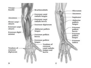
Anatomy • Largest sesamoid bone • Thick articular cartilage proximally • Articular surface divided into medial and lateral facets by longitudinal ridge • Distal pole nonarticular
Anatomy • Patellar Retinaculum • Longitudinal tendinous fibers • Patellofemoral ligaments • Blood Supply • Primarily derived from geniculate arteries
Biomechanics • The patella undergoes approximately 7 cm of translation from full flexion to extension • Only 13-38% of the patellar surface is in contact with the femur throughout its range of motion
Biomechanics • The patella increases the moment arm about the knee • Contributes up to 30% increase in force with extension • Patella withstands compressive forces greater than 7X body weight with squatting
Biomechanics • Twice as much torque is needed to extend the knee the final 15 degrees than to extend from a fully flexed position to 15 degrees of flexion
History • Direct blow to the anterior knee (dashboard injury) • Fall from height • Rapid knee flexion with quadriceps resistance
Physical Examination • Pain, swelling, contusions, lacerations and/or abrasions at the site of injury • Palpable defect • Assessment of ability to extend the knee against gravity or maintain the knee in full extension against gravity
Radiographic Evaluation • AP & Lateral • Patella alta or baja • Note fracture pattern • Articular step-off, diastasis • Special views • Axial or sunrise • CT Scan -Occult fractures
Radiographic Evaluation • Bipartite Patella • Obtain bilateral views • Often involves superolateral corner • Accessory ossification center
Etiology • Allows prediction of outcome • Direct trauma • Dashboard injury • Increasing cases with penetrating trauma • Often with comminution and articular damage • Indirect trauma • Violent flexion directed through the extensor mechanism against a contracted quadriceps • Results in simple, transverse fractures
Classification • Allows prediction of treatment • Types • Transverse • Marginal • Vertical • Comminuted • Osteochondral
Nonoperative Treatment • Indicated for nondisplaced fractures • <2mm of articular stepoff and <3mm of diastasis with an intact extensor mechanism • May also be considered for minimally displaced fractures in the elderly • Patients with a extensive medical comorbidities
Nonoperative Treatment • Long leg cylinder cast for 4-6 weeks • May consider a knee immobilizer for the elderly • Immediate weightbearing as tolerated • Rehabilitation includes range of motion exercises with gradual quadriceps strengthening
Operative Treatment • Goals • Preserve extensor function • Restore articular congruency • Preoperative Setup • Tourniquet • Prior to inflation, gently flex the knee • Approach • Longitudinal midline incision recommended • Transverse approach alternative • Consider future surgeries!
Operative Techniques • Modified tension band wiring • Lag-screw fixation • Cannulated lag-screw with tension band • Partial patellectomy • Patellectomy
Modified Tension Band Wiring • Transverse, noncomminuted fractures • After reduction, fracture is fixed with two parallel, 1.6mm Kirschner wires placed perpendicular to the fracture • 18 gauge wire passed behind proximally and distally
Modified Tension Band Wiring • Wire converts anterior distractive forces to compressive forces at the articular surface • Two twists are placed on opposite sides of the wire • Tighten simultaneously to achieve symmetric tension • Repair any retinacular tears
Lag-Screw Fixation • Indicated for stabilization of comminuted fragments in conjunction with tension band wiring or cerclage wires • May also be used as an alternative to tension band wiring for transverse or vertical fractures
Lag-Screw Fixation • Contraindicated for extensive comminution and osteopenic bone • Small secondary fractures may be stabilized with 2.7mm or 3.5mm cortical screws • Transverse or vertical fractures require 3.5mm or 4.5mm cortical screws • Retrograde insertion of screws may be technically easier
Cannulated Lag-Screw With Tension Band • Fully threaded screws placed with a lag technique • Wire through screws and across anterior patella in figure of eight tension band
Cannulated Lag-Screw With Tension Band • Most stable construct • Screws and tension band wire combination eliminates both possible separation seen at the fracture site with modified tension band and screw failure due to excessive three point bending
Gosal et al Injury 2001 Wire v. #5 Ethibond 37 patients Reoperation 38% wire group vs. 6% Infection 3 pts wire group vs. 0 Patel et al, Injury 2000 McGreal et al, J Med Eng Tech, 1999 Cadaveric models Quality and stability of fixation comparable to wire Conclude suture an acceptable alternative Suture vs. Wire Tension Band
Partial Patellectomy • Indicated for fractures involving extensive comminution not amenable to fixation • Larger fragments repaired with screws to preserve maximum cartilage • Smaller fragments excised • Usually involving the distal pole
Partial Patellectomy • Tendon is attached to fragment with nonabsorbable suture passed through drill holes in the fragment • Drill holes should be near the articular surface to prevent tilting of the tendon and minimize articular step-off • Watch for patellar tilt! • Load sharing wire passed through drill holes in the tibial tubercle and patella may be used to protect the repair and facilitate early range of motion
Total Patellectomy • Indicated for displaced, comminuted fractures not amenable to reconstruction • Bone fragments sharply dissected • Defect may be repaired through a variety of techniques • Usually results in extensor lag and loss of strength
Postoperative Management • Immobilization with knee brace • Immediate WBAT • Early range of motion • Based on intraoperative assessment of repair • Active flexion with passive extension • Quadriceps strengthening • Begun when there is radiographic evidence of healing, usually around 6 weeks
Knee Stiffness Most common complication Infection Rare, depends on soft tissue compromise Loss of Fixation Hardware failure in up to 20% of cases Osteoarthritis May result from articular damage or incongruity Nonunion < 1% with surgical repair Painful hardware Removal required in approximately 15% Complications
Extensor Tendon Ruptures • Patellar and quadriceps tendon ruptures are uncommon injuries • Patients are typically males in their 30’s or 40’s • Patellar < 40 yo • Quadriceps > 40 yo • Fall, sports, MVA
Quadriceps Tendon Rupture • Typically occurs in patients > 40 years old • Usually 0-2 cm above the superior pole • Level often associated with age • Rupture occurs at the bone-tendon junction in majority of patients > 40 years old • Rupture occurs at midsubstance in majority of patients < 40 years old
Quadriceps Tendon Ruptures • Risk Factors • Chronic tendonitis • Anabolic steroid use • Local steroid injection • Inflammatory arthropathy • Chronic renal failure • Systemic disease
History • Sensation of a sudden pop while stressing the extensor mechanism • Pain at the site of injury • Inability/difficulty weightbearing
Physical Exam • Effusion • Tenderness at the upper pole • Palpable defect above superior pole • Loss of extension • With partial tears, extension will be intact
Radiographic Evaluation X-ray- AP, Lateral, and Tangential (Sunrise, Merchant) Distal displacement of the patella MRI Useful when diagnosis is unclear Treatment Nonoperative Partial tears and strains Operative For complete ruptures Quadriceps Tendon Rupture
Operative Treatment • Reapproximation of tendon to bone using nonabsorbable sutures with tears at the muscultendonous junction • Locking stitch (Bunnel, Krakow) with No. 5 ethibond passed through vertical bone tunnels • Repair tendon close to articular surface to avoid patellar tilting
Operative Treatment • Midsubstance tears may undergo end-to-end repair after edges are freshened and slightly overlapped • May benefit from reinforcement from distally based partial thickness quadriceps tendon turned down across the repair site (Scuderi Technique)
Treatment • Chronic tears may require a V-Y advancement of a retracted quadriceps tendon (Codivilla V-Y-plasty Technique)
Postoperative Management • Knee immobilizer or cylinder cast for 5-6 weeks • Immediate vs. delayed (3 weeks) weightbearing as tolerated • At 2-3 weeks, hinged knee brace starting with 45 degrees active range of motion with 10-15 degrees of progression each week
Complications • Rerupture • Persistent quadriceps atrophy/weakness • Loss of motion • Infection
Patellar Tendon Rupture • Less common than quadriceps tendon rupture • Associated with degenerative changes of the tendon • Rupture often occurs at inferior pole insertion site
Patellar Tendon Rupture • Risk Factors • Rheumatoid • Systemic Lupus Erythematosus • Diabetes • Chronic Renal Failure • Systemic Corticosteroid Therapy • Local Steroid Injection • Chronic patellar tendonitis
Anatomy • Patellar tendon • Averages 4 mm thick but widens to 5-6 mm at the tibial tubercle insertion • Merges with the medial and lateral retinaculum • 90% type I collagen
Blood Supply • Fat pad vessels supply posterior aspect of tendon via inferior medial and lateral geniculate arteries • Retinacular vessels supply anterior portion of tendon via the inferior medial geniculate and recurrent tibial arteries • Proximal and distal insertion areas are relatively avascular and subsequently are a common site of rupture
Biomechanics • Greatest forces are at 60 degrees of flexion • 3-4 times greater strain are at the insertions compared to the midsubstance prior to failure • Forces through the patellar tendon are 3.2 times body weight while climbing stairs
History • Often a report of forceful quadriceps contraction against a flexed knee • May experience and audible “pop” • Inability to weightbear or extend the knee
Physical Examination • Palpable defect • Hemarthrosis • Painful passive knee flexion • Partial or complete loss of active extension • High riding patella on radiographs
Radiographic Evaluation • AP and Lateral X-ray • Patella alta seen on lateral view • Patella superior to Blumensaat’s line • Ultrasonagraphy • Effective means to determine continuity of tendon • Operator and reader dependant • MRI • Effective means to assess patellar tendon, especially if other intraarticular or soft tissue injuries are suspected • Relatively high cost
Classification • No widely accepted means of classification • Can be categorized by: • Location of tear • Proximal insertion most common • Timing between injury and surgery • Most important factor for prognosis • Acute- within two weeks • Chronic- greater than two weeks
Treatment • Surgical treatment is required for restoration of the extensor mechanism • Repairs categorized as early or delayed
Early Repair • Better overall outcome • Primary repair of the tendon • Surgical approach is through a midline incision • Incise just lateral to tibial tubercle as skin thicker with better blood supply to decrease wound complications • Patellar tendon rupture and retinacular tears are exposed