Download
1 / 12
140 likes | 387 Views
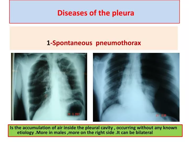
Diseases of the pleura. 1 -Spontaneous pneumothorax. Is the accumulation of air inside the pleural cavity , occurring without any known etiology .More in males ,more on the right side .It can be bilateral .

E N D
Diseases of the pleura 1-Spontaneous pneumothorax Is the accumulation of air inside the pleural cavity , occurring without any known etiology .More in males ,more on the right side .It can be bilateral
Causes 1- Ruptured pulmonary bleb.2-Ruptured of a cystic defect in the pleura.3-Teared visceral pleura 4-No cause can be demonstrated in (15-20%).Complications:-1-pleural effusion2-empyema 3-tension pneumothorax which leads to mediastinal shift &circulatory collapse.4-Respiratory failure in elderly patient with COAD . • Treatment :- • 1-Bed rest ,O2 administration &observation in limited pneumothorax. • 2-Aspiration • 3-Chest tube (thoracostomy tube or ICD intercostal drain in a safety triangle which is bounded by pectoralis muscle anteriorly &lattismus muscle posteriorly and the superior border of the nipple.in the fifth intercostal space just anterior to the mid axillary line to avoid the long thoracic nerve . • 4-bronchoscopy is indicated if the lung fail to expand • 5-Chemical pleurodesis.by injecting sclerosing agent as Tetra cycline • 6-Surgery pleurectomy by thoracotomy or thoracoscopically if the lung fail to re expand
2-Spontaneous haemothorax • Is the presence of blood inside the pleural cavity • Causes:- • 1-pulmonary causes ----------TB , AV malformation • 2-pleural causes -----------torn vascular adhesion • 3-pulmonary malignancy ….primary or metastatic • 4-blood dyscrasia ……………..hemophilia • 5-abdominalpathology ……….. haemo peritoneum • 6-thoracic causes ………ruptured great vessels
Clinical featuresdyspnea , chest pain ,syncopesigns of hypovolaemic shock blood inside the pleural cavity may leads to deposition of fibrin on the pleural surface leading to fibrosis (trapped lung syndrome) . • Treatment • 1-Resuscitation • 2-Tube thoracostomy • 3-May needs thoracotomy if excessive bleeding • initial bleeding more than 1.5 liter • Or continuous bleeding more than 200 ml/hour for more than 4 hours
3-Chylo –thorax Is the presence of lymph in the pleural space Causes A-Congenital atresia of the thoracic duct , birth trauma B-Traumatic C-Neoplastic malignancy D-Infection TB Diagnosis milky pleural effusion that does not clot and contains fat , fat soluble vitamins & antibodies Treatment 1-Conservative consists of insertion of tube thoracostomy to drain the effusion , correction of the fluid and electrolytes with nutritional supplement. 2-Surgery consists of ligation of the thoracic duct if the effusion continues for more than two weeks .
4-Pleural effusion • Is the accumulation of fluid in the pleural space excessive transudation or exudation of the interstitial fluid from the pleural surface. It is signfrom ify pleural or systemic disease . • Its effect depends on its size (mild , moderate or massive ) & the state of the underlying lung .It is classified as transudate when the protein content is less than 3g/100ml, or exudates when protein content is more than 3 gm /100ml.Clinically patients will present with dyspnea & pleuritic chest pain • Radio logically (concave meniscus sign) • Transudate as in CHF • Exudate as in malignancy • Treatment :-1-aspiration (thoracentesis) 2-tube thoracostomy • 3-chemicalpleurodesis 4-pleuectomy to remove the pleura to stop theeffusion.
5-Empyaema • Is the accumulation of pus in the pleural space , it passes into three stages • 1-Acute phase with the clinical manifestation of fever & toxicity . • 2-Transitional phase with the increased turbidity of the fluid & decrease the size of the lung . • 3-Chronic phase with the pleural thickening ,decrease amount of the fluid &the development of the trapped lung syndrome .
Tube ThoracostomyTube thoracostomy or Chest Tube or ICD(Intercostal drain)Is a flexible hollow plastic tube that is inserted through the chest wall into the pleural space and connected to a bedside drainage container Indications:- 1-Pnemothorax
3-Hydro thorax4-Chylothorax 5-Thoracic Operation (Tube Thoracostomy without trocar ) .On the lung or Mediastinum Or The esophagus6-Postoperative (Collection or Infected space ).7-malignant Effusiondrainage and giving medicationthrough it.