Download
1 / 1
10 likes | 141 Views
Longitudinal fNIRS Stroop Study of Adult Traumatic Brain Injured Patients in Post-Acute Treatment. 1 Pate Rehabilitation, 2 The University of Texas Arlington. Introduction: Traumatic brain injury (TBI) and stroke are the leading causes of adult disability and death

E N D
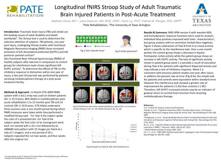
Longitudinal fNIRS Stroop Study of Adult Traumatic Brain Injured Patients in Post-Acute Treatment 1Pate Rehabilitation, 2The University of Texas Arlington Introduction: Traumatic brain injury (TBI) and stroke are the leading causes of adult disability and death worldwide1. The Stroop test is used to determine the selective attention of subjects. TBI patients, one year post injury, undergoing Stroop studies with functional Magnetic Resonance Imaging (fMRI) show increased activation of left dorsolateral prefrontal (DLPFC) and left posterior parietal cortices2. Also functional Near-Infrared Spectroscopy (fNIRS) of healthy subjects after exercise in comparison to control groups for interference tasks shows significant left DLPFC activity3. To determine the effects of TBI on the inhibitory response to distraction within one year of injury, a two part Stroop task was performed by patients receiving multidisciplinary therapy at a post-acute treatment facility. Results & Summary: NIRS-SPM version 4 with wavelet-MDL and hemodynamic response functions were used for analysis. Individual false positives improved with Euler characteristics4, but group analysis with no correction limited false negatives. Figure 3shows subtraction of Task B from A to reveal activity which is specific to the interference task. Over a one month period, the control group shows a decrease in bipolar frontopolar cortex activity while the patient group shows an increase in left DLPFC activity. The lack of significant activity shown in patient group week 1 is possibly a result of saturation during Task A for patients with significant dispersed activity and may indicate a lack of inhibitory response. Week 5 is consistent with previous patient studies one year after injury. In addition the percent rate of error (Fig 4) for the simple task for patients and controls were equivalent within standard error. However the interference task showed a greater rate of improvement for patients (-5.63) than controls (-1.98.) Therefore, left DLPFC increased activity may be an indicator of gradual return to normal brain function from receiving multidisciplinary therapy. Controls Patients 1st Week 3rd Week 5th Week Methods & Approach : A Hitachi ETG-4000 fNIRS system with a 3x11 array was used on Sixteen patients (41 +/-15.0 years, 86% Male) in multidisciplinary post-acute rehabilitation 2 to 13 months post TBI and 14 controls (38 +/-10.8 years, 57% Male) underwent three sessions over a one month period during which fNIRS measures were taken while they performed a modified Stroop task. For Task A the subject spoke the color of a presented dot. For Task B the subject spoke the font color of an incongruent word. Each run started with a 10 s rest followed by an ABBABA task pattern with 25 images per block at a rate of 1 image/s, and a rest period of 30 s. Subjects repeated the run two weeks, and four weeks after the original run. Matthew Cloud, BS1,2; Jana Downum, MA, BCB, CBIS1; Hanli Liu, PhD2; Patrick M. Plenger, PhD, ABPP1 1st Week 3rd Week 5th Week References: 1U.S. Centers for Disease Control and Prevention 2Soeda, A, et al., “Cognitive Impairment After Traumatic Brian Injury: a Functional Magnetic Resonance Imaging study using the Stroop Task,” Neuroradiology 47:501-506, 2005. 3Yanagisawa, H, et al., “Acute Moderate Exercise Elicits Increased Dorsolateral Prefrontal Activation and Improves Cognitive Performance with Stroop Test,” NeuroImage50:1702-1710, 2010. 4Li, H., et al., “Lipschitz Killing curvature based expected Euler characteristics for p-value correction in fNIRS,” J. Neurosci. Meth. 204, 61-67, 2012. 5 Hitachi Medical Systems Europe 1994, 2012 6Ye, J. C., et al., “NIRS-SPM: Statistical parametric mapping for near-infrared spectroscopy,” NeuroImage 44, 428-447, 2009. Fig 3) Interference task minus simple task (B-A) p-value=0.05, no correction; Control Group (n=14, 13, 13); Patient Group (n=16, 14, 16) Fig 4) Stroop Error Rate by Task with Standard Error Fig 1) 3x11 Optode Array5 Fig 2) Probe placement5 For presentation at the 2ndBiennual Functional Near-Infrared Spectroscopy Conference Funding: Pate Rehabilitation Endeavors, Inc.