Download
1 / 73
920 likes | 1.6k Views
Acute Respiratory Distress Syndrome. Dr. Vanya Chugh. University College of Medical Sciences & GTB Hospital, Delhi. Timeline. In 1967 – Ashbaugh, Bigelow, Petty, Levine - described Acute Respiratory Distress Syndrome in adults

E N D
Acute Respiratory Distress Syndrome Dr. VanyaChugh University College of Medical Sciences & GTB Hospital, Delhi
Timeline • In 1967 – Ashbaugh, Bigelow, Petty, Levine - described Acute Respiratory Distress Syndrome in adults • In 1971, Petty and Ashbaugh modified its name from ‘acute’ to ‘adult’ Respiratory Distress Syndrome; to differentiate it from its newborn counterpart • In 1974, Webb and Tierney confirmed the existence of ventilator associated lung injury • In 1990, Hickling et al introduced the concept of permissive hypercapnia
Timeline • In 1992, American European Consensus Conference (AECC) gave standardized definition for ARDS • In 1997, Tremblay et al introduced the concept of biotrauma • In 1998, Amato et al, conducted RCT - decrease in mortality using low tidal volume ventilation and high PEEP (open lung strategy) • In 2000, ARDS network trial demonstrated the benefits of low tidal volume and PEEP ventilation
Definitions of ARDS Ashbaugh and colleagues, 1967 • Severe dyspnea • Tachypnea • Cyanosis refractory to oxygen therapy • Decreased pulmonary compliance • Diffuse alveolar infiltrates on chest radiograph. • Loosely defined criteria • Definition of hypoxemia inconsistent
Murray & Mathay Lung Injury Score(1988) Chest Radiology findings Score No alveolar consolidation 0 One quadrant 1 Two quadrant 2 Three quadrant 3 Four quadrant 4 Oxygenation status (Hypoxemia Score) PaO2 / FiO2 > 300 mmHg 0 225-299 mmHg 1 175-224 mmHg 2 100-174 mmHg 3 < 100 mmHg 4
Pulmonary compliance Score Compliance (ml/cmH2O) > 80 0 60-79 1 40-59 2 20-39 3 < 19 4 PEEP settings (when ventilated) PEEP (cmH2O) < 5 0 6-8 1 9-11 2 12-14 3 > 15 4 Acute lung injuries assessed by dividing sum by 4 0 points = No pulmonary injury 0.1-2.5 points = Mild to moderate > 2.5 points = Severe (ARDS)
Murray & Mathay Lung Injury Score Advantages : • Ventilatory settings included Disadvantage : • Complex • Lacks prospective validity
Bernard and colleagues, 1992 (American European Consensus conference definition) A three-criteria system including chest radiograph, oxygenation score, and exclusion of cardiogenic causes: • Acute onset, bilateral infiltrates on chest radiography, • Acute lung injury ~ PaO2/FIO2 ≤ 300 ARDS subset~ PaO2/FIO2 ≤ 200 • Pulmonary-artery wedge pressure of <18 mm Hg or the absence of clinical evidence of left atrial hypertension
Bernard and colleagues, 1992 (American European Consensus conference definition) Problems • Acute onset : arbitrary; <1 week • Bilateral infiltrates: inter observer variation, b/l pneumonia, atelectasis, cardiogenic pulmonary edema • PAOP of <18 mm Hg /absence of clinical evidence of left atrial hypertension : PAOP: poor estimate of PVH, falsely raised with high airway pressures • Acute lung injury present if PaO2/FIO2 is 300 : new and arbitrary value
Delphi definition (2005) of ARDS Diagnosis : 1- 4 present with 5a and/or 5b1. PaO2/FiO2 ratio ≤ 200 on PEEP ≥ 10.2. Bilateral airspace disease : ≥ 2 quadrants, frontal chest X-ray 3. Onset within 72 hours.4. No clinical evidence/subjective finding of CHF (including use of PA catheter and/or echo if clinically indicated) 5a. Static respiratory compliance < 50ml/cm H2O (patient sedated, TV 8ml/kg, PEEP ≥ 10.5b. Presence of direct or indirect risk factor associated with lung injury.
Delphi definition of ARDS contd. • Airspace disease – presence of one or more of the following- • Air brochogram • Acinar shadows • Coalescence of acinar shadows • Silhoutte sign • Specificity : delphi = LIS > AECC • Sensitivity : delphi = LIS = AECC • Delphi criteria provisional, need further testing
Synonyms of ARDS • Shock lung • Pump lung • Traumatic wet lung • Post traumatic atelectasis • Adult hyaline membrane disease • Progressive respiratory distress • Acute respiratory insufficiency syndrome • Haemorrhagicatelectasis • Hypoxic hyperventilation • Postperfusion lung • Oxygen toxicity lung • Wet lung • White lung • Transplant lung • Da Nang lung • Diffuse alveolar injury • Acute diffuse lung injury • Noncardiogenic pulmonary edema. • Progressive pulmonary consolidation
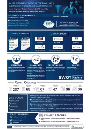
Epidemiology of ARDS • Difficult to estimate • Lack of standardization of the definition • Difference in methodology • KCLIP study (1999-2000) done on ARDS patients as per AECC criteria estimated - - incidence of ALI – 78.9/lakh person years - mortality rate – 38.5- 41.1%
Precipitating Factors Direct Lung Injury • Pneumonia • Aspiration of gastric contents • Pulmonary contusion • Near-drowning • Toxic inhalation injury Indirect Lung Injury • Sepsis • Severe trauma Multiple bone fractures Flail chest Head trauma Burns • Multiple transfusions • Drug overdose • Pancreatitis • Post-cardiopulmonary bypass
Differential risk factors • Chronic alcohol abuse • Absence of DM • Age • Gender • Severity of illness – APACHE score • Excessive blood transfusion • Cigarette smoking
Pathophysiology in ARDS Based on the histological appearance - Exudative phase (0-4 days) • Alveolar and interstitial edema • Capillary congestion • Destruction of type I alveolar cells • Early hyaline membrane formation Proliferative Phase (3-10 days) • Increased type II alveolar cells • Cellular infiltration of alveolar septum • Organisation of hyaline membranes Fibrotic Phase (>10 days) • Fibrosis of hyaline membranes and alveolar septum • Alveolar duct fibrosis
Pathology in ARDS Mechanisms in early phase - • Release of inflammatory cytokines – TNF alpha, IL- 1,6,8 • Failure of alveolar edema clearance, epithelial and endothelial damage • Increased permeability of alveolo – capillary membrane • Neutrophil migration and oxidative stress • Procoagulant shift – fibrin deposition • Surfactant dysfunction Mechanism in late (repair) phase – • Fibroproliferation -TGF beta, MMPs, thombospondin, plasmin, ROS • Remodelling - matrix and cell surface proteoglycans, MMP, imbalance of coagulation and fibrinolysis.
D/D : Hydrostatic pulmonary edema • PCWP ≥ 18 mmHg • Causes : • Cardiogenic – LVF (eg. MI, myocarditis) cardiac valvular disease (aortic, mitral) • Vascular – systemic HTN, pulmonary embolism • Volume overload - excessive iv fluids, renal failure
Cardiogenic vs Non-cardiogenic edema Cardiogenic Non-cardiogenic 1. Prior h/o cardiac disease Absence of heart disease 2.Third heart sound No third heart sound 3. Cardiomegaly Normal sized heart 4. Infiltrates : Central distribution Peripheral distribution 5. Widening of vascular pedicle No widening of vascular pedicle ( ↑ width of mediastinum) 6. PA wedge pressure N or PA wedge pressure 7. Positive fluid balance Negative fluid balance
Management • Treatment of the precipitating cause • Mechanical ventilation – • Core ventilator management - protective lung ventilation strategy - role of ‘open lung approach’ • Adjuncts to core ventilation - • Fluid restriction • Permissive hypercapnia • Prone positioning • Recruitment maneuvers
Management contd. • Non conventional/Salvage interventions • High frequency ventilation • Airway pressure release ventilation • Tracheal gas insufflation • Inverse ratio ventilation • Inhaled nitric oxide • Inhaled prostacyclin • Corticosteroids • Surfactant administration • Liquid ventilation • Extracorporeal membrane oxygenation • Supportive therapy – nutrition, prevention of infection
Concept of VALI • Mechanical ventilation - Basic care in critically ill ICU patients • May cause or worsen lung injury – ventilator induced/associated lung injury • Components – • Barotrauma • Volutrauma • Atelectrauma • Biotrauma
Concept of ‘baby lung’ • Put forward by Gattinoni and colleagues first in 1987 • Lung injury in ARDS - non homogenous, basal • Edema and consolidation > dependent lung regions - ↑ density of dorsal regions • Aerated ventral regions – ‘baby lung’ (300-500gms) – high compliance • Ventilation of baby lung with normal tidal volumes and pressures – alveolar over distension – injury to functional lung tissue
Management Lung protective ventilation ARDS network protocol • Goals • Oxygenation : PaO2 55-80 mmHg, or SpO2 88 – 94% (excluding pregnancy, intracranial hypertension or stroke where SaO2 goal>94%) • Ventilation : • Tidal volume : 4-6 ml/kg ideal body weight • Plateau pressure : <30cmH2O • Ph: 7.25-7.35 • I:E ratio of 1:1 – 1:3
Management contd. Oxygenation • Initially high Fio2 given (1.0) to correct hypoxia • Fio2 and PEEP adjusted to the lowest level compatible with the oxygenation goals • Fio2 and PEEP adjusted in the following fixed combinations {fio2/PEEP(mmHg)} FIO2 PEEP
Management contd Initial ventilator set up and adjustments STEP 1- Calculation of ideal body weight(IBW): • For males, IBW(kg) = 50+2.3{height(inch)– 60} Or IBW(kg)=50 + 0.91{height(cm)–152.4} • For females, IBW(kg) = 45.5+2.3{height(inch)– 60} Or IBW(kg)=45.5 + 0.91{height(cm)–152.4}
Management contd STEP 2 - Volume assist control selected as ventilator mode • Initial tidal volume (TV) set at 8ml/kg IBW • TV reduced by 1ml/kg IBW 2 hourly until TV = 6ml/kg IBW • Initial ventilator rate set to maintain baseline minute ventilation( not >35/min) • TV and respiratory rate adjusted to achieve the pH and plateau pressure goals • Inspiratory flow rate set above patients demand (usually >80L/min)
Open Lung Approach • Introduced by Amato et al in 1998 – use of low tidal volume + high PEEP+ recruitment (Open lung strategy) – reduce mortality in ARDS • Maintaining inflation & deflation between 2 inflection points during entire respiratory cycle • Ventilatory settings - PEEP >Pflex & TV reduced so that Pplat < UIP • Advantages- avoids repetitive opening and closing of alveoli (VALI) - minimizes shear injury
Management • Treatment of the precipitating cause • Mechanical ventilation – • Core ventilator management – protective lung ventilation strategy role of ‘open lung approach’ • Adjuncts to core ventilation – • Fluid restriction • Permissive hypercapnia • Prone positioning • Recruitment maneuvers
Fluid restriction in ARDS • Rationale – alveolar flooding depends on : • Capillary hydrostatic pressure • Oncotic pressure • Alveolar–capillary permeability • Capillary permeability increased in ARDS • ↓ hydrostatic pressure and ↑ oncotic pressure may help.
Fluid therapy in ARDS Recommended : • Central venous pressure guided therapy – 10-14 mmHg ( ARDS Network Trial 2003) • Restricted fluid intake • Increased urine output – Diuretics or RRT Not recommended : • Vasodilators • Albumin
Management • Treatment of the precipitating cause • Mechanical ventilation – • Core ventilator management - protective lung ventilation strategy - role of ‘open lung approach’ • Adjuncts to core ventilation – • Fluid restriction • Permissive hypercapnia • Prone positioning • Recruitment maneuvers
Permissive Hypercapnia • Hickling and colleagues 1990 • “Degree of hypercapnia permitted in patients subjected to lower tidal volumes” • Upper limit – not defined; >100 mmHg avoided • Advantages • Increased surfactant secretion (animal models) – improved V/Q match, oxygenation (improved compliance) • Increased cardiac output and oxygen delivery (sympathoadrenal effects predominate over cardiodepressant effects) • Increased cerebral blood flow and tissue oxygenation
Permissive Hypercapnia • Concerns • Increase in pulmonary vascular resistance • Impaired diaphragmatic function (impairs afferent transmission) • Decrease in cardiac contractility • Raised intracranial tension • Individualize and treat
Management • Treatment of the precipitating cause • Mechanical ventilation – • Core ventilator management - protective lung ventilation strategy - role of ‘open lung approach’ • Adjuncts to core ventilation – • Fluid restriction • Permissive hypercapnia • Prone positioning • Recruitment maneuvers
Prone Position Ventilation • First suggested by Piehl and Brown in 1976 • Offers improved oxygenation by: • Increased FRC • Change in regional diaphragm motion • Distribution of perfusion • Better clearance of secretions
Prone Position Ventilation • Sud and colleagues conducted – meta-analysis of 13 RCTs (1559 patients) on supine and prone position ventilation in ARDS/ALI patients • Median MV of 12 hours ( 4-24hrs) for 4 days( 1-10 days) • Conclusion -cannot be recommended for routine Mx -no evidence of improved survival • Gattinoni et al suggested no overall reduction in mortality except in very sick patients ( SAPS II Score >50) • No decrease in ventilator associated pneumonia
Problems of prone position • Facial edema • Airway obstruction • Difficulties with enteral feeding • Transitory decrease in oxygen saturation • Hypotension & Arrhythmias • Vascular and nerve compression • Loss of venous accesses and probes • Loss of chest drain and catheters • Accidental extubation • Apical atelectasis d/t incorrect positioning of the tracheal tube • Increased need for sedation
Management • Treatment of the precipitating cause • Mechanical ventilation – • Core ventilator management - protective lung ventilation strategy - role of ‘open lung approach’ • Adjuncts to core ventilation – • Fluid restriction • Permissive hypercapnia • Prone positioning • Recruitment maneuvers
Recruitment maneuvers • High pressure inflation maneuver aimed at temporarily raising the transpulmonary pressure above levels typically obtained with mechanical ventilation • Types – Elevated sustained pressures : 40 cm H2O for 40 seconds Sigh breaths : ↑ tidal volume / PEEP for one or several breaths Extended sigh breath : VCV with PEEP well above LIP for a longer time • More effective in early ALI and those with more homogenous disease; atelectasis > consolidation.
Recruitment maneuvers Adverse effects • Hypotension • Barotrauma • Raised ICP • Haemodynamic instability
Management contd. • Non conventional/Salvage interventions • High frequency ventilation • Airway pressure release ventilation • Tracheal gas insufflation • Inverse ratio ventilation • Inhaled nitric oxide • Inhaled prostacyclin • Corticosteroids • Surfactant administration • Liquid ventilation • Extracorporeal membrane oxygenation • Supportive therapy – nutrition, prevention of infection
High Frequency Ventilation • Mechanical ventilatory support using higher than normal breathing frequencies • Smaller tidal pressure swings (within inflection points) along with apt mpaw • Smaller tidal volumes and higher mean pressure utilized for lung protection • Special ventilators required • Types - High Frequency Jet Ventilation (HFJV) High Frequency Oscillatory Ventilation (HFOV)
HFV HFJV • A nozzle/injector creates high velocity ‘jet’ of gas directed into the lung • Injectors – 1-3mm diameter • Expiration is passive • Frequencies available – upto 600 breaths/min • Available for neonatal and paediatric use only HFOV • Characterized by rapid oscillations of a diaphragm (at 3 to 10 hertz i.e 180 to 160 breaths/min) driven by a piston pump • Frequencies available – 300-3000 breaths/min • Expiration is also active – risk of air trapping minimal
HFV contd Advantages • Better oxygenation and ventilation • Aids lung recruitment (high mpaw) • Reduces oxygen toxicity (high mpaw) • Minimizes VILI Disadvantages • Delivered tidal volumes difficult to monitor • Deep sedation and/or paralysis required • Inadequate humidification • Direct physical airway damage
Airway Pressure Release Ventilation • Alternative mode of ventilation that applies a form of CPAP that is released periodically, augmenting CO2 release. • Pressure limited, time cycled mode • Permits spontaneous ventilation throughout the respiratory cycle • Based on the ‘open lung’ concept – maximize and maintain recruitment throughout the respiratory cycle
APRV contd • Uses 2 airway pressures – P high and P low; 2 set time periods – T high and T low, usually T high>T low • P high is set above the closing pressure of recruitable alveoli (lower inflection point) • Set T high maintains the P high for several seconds • T low helps remove CO2