Download
1 / 22
220 likes | 238 Views
Skeletal System Introduction Page 22. I. What is the Skeletal System?. The skeletal system is the organ system composed of bones, joints, cartilage, and ligaments that allow us to…. A. Functions of the Skeletal System: 1. Supports the body 2. Protects and cradles vital organs

E N D
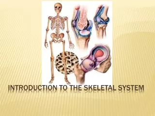
I. What is the Skeletal System? • The skeletal system is the organ system composed of bones, joints, cartilage, and ligaments that allow us to…
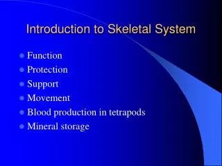
A. Functions of the Skeletal System: • 1. Supports the body • 2. Protects and cradles vital organs • 3. Movement: works with muscles to move the body • 4. Storage of minerals (calcium and phosphorus) and growth factors • 5. Blood Cell Formationin bone marrow • 6. Triglyceride (fat) storage
Adults have ~206-208 bones • Babies have ~275 bones • Why the difference? • Many of the babies bones are very soft and will fuse together as they age (think soft spot) • B. Bone Features: • Living organ • Very hard (calcium salts) • Light weight • Ability to resist tension and forces (collagen fibers)
C. Divisions of the Skeletal System • Axial Skeleton: bones of the skull, vertebral column, and rib cage • Appendicular Skeleton: bones of the upper and lower limbs, shoulder, and hip.
Axial Skeleton Appendicular Skeleton Clavicle (collarbone) Scapula (shoulder blade) Coxal (pelvic girdle) Humerus (arm) Radius, ulna (forearm) Carpals (wrist) Metacarpals (hand) Phalanges (fingers, toes) Femur (thigh) Tibia, fibula (leg) Tarsal, metatarsals (foot) Calcaneus (heel) Patella (knee) • Cranium (skull) • Mandible (jaw) • Vertebral column (spine) • Cervical vertebrae • Thoracic vertebrae • Lumbar vertebrae • Sacrum • Coccyx • Sternum (breastbone) • Ribs
II. How are bones classified? • The bones of the skeletal system differ greatly in size and shape. However, they are similar in structure, development, and function. • Bones are classified according to their shapes- long, short, flat, or irregular.
A. Bone Classification • 1. Long Bones: • Longer than they are wide • Examples: Femur (upper leg), Humerus (upper arm), metacarpals (palm)
2. Short Bones: • Cube-shaped bones with roughly equal lengths and widths. • Examples: Bones of the carpals (wrist) and tarsals (ankle) • Bones that form within tendons= sesamoid bones (like the patella)
3. Flat Bones: • Bones that are thin, flat, and slightly curved • Examples: sternum, most skull bones, and scapula (shoulder blade)
4. Irregular Bones: • Bones with complicated shapes. • Examples: vertebrae (around spine), hip bones, and many facial bones.
What type of bone? Humerus Sternum Long Bone Flat Bone Tarsals= ankle bones Vertebrae Short Bone Irregular Bone
B. Structure of a Long Bone • Long Bone Anatomy (Humerus) • 1. Epiphysis= end of bone (proximal or distal) • 2. Diaphysis= long shaft of the bone, between the epiphyses • 3. Metaphysis= epiphyseal growth plate • 4. Articular cartilage • Hyaline cartilage coats outer surface of epiphysis (end)
5. Periosteum • Tough outer covering of the bone; dense connective tissue; where blood vessels and nerve fibers connect to bone • 6. Medullary cavity • Hollow chamber in diaphysis • 7. Endosteum • Thin layer of cells that lines the medullary cavity
Growth plates= Metaphysis Articular cartilage Proximal Epiphysis Endosteum Diaphysis Medullary Cavity Periosteum Distal Epiphysis
III. Histology of Bone Tissue • A. Bone Cells: • 1. Osteoblasts: bone-forming cells; make extracellular matrix of bone tissue • 2. Osteocytes:mature bone cells (don’t divide through mitosis); exchange nutrients and waste with blood • 3. Osteogenic cells: undergo cell division (mitosis) and develop into osteoblasts
B. Types of Bone Tissue: • 1. Compact Bone: • Tightly packed bone tissue with no visible open spaces (extremely hard and resists bending). • Makes up wall mainly of Diaphysis • Blood vessels go through to cells= Haversian canals • 2. Spongy Bone: • Thin layers of bone tissue with open spaces= trabeculae • Provides most of the strength and support because it absorbs shock easily • Makes up mainly epiphyses
Spongy Bone Compact Bone
3. Red Marrow: • Fills middle of long bones • Produces red and white blood cells • 4. Yellow Marrow: • Fills the very center of long bones (medullary cavity) • Stores fat(made up of mostly fat cells)
Draw and Label Bone Tissue: Spongy Bone Red Marrow Yellow Marrow Compact Bone