Download

1 / 31
320 likes | 722 Views
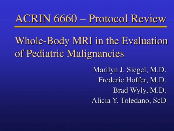
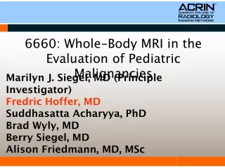
Whole-Body MRI in the Evaluation of Pediatric Malignancies. Marilyn J. Siegel, M.D. Frederic Hoffer, M.D. Brad Wyly, M.D. Alicia Y. Toledano, ScD. ACRIN 6660 – Protocol Review. Aims. Primary Aim

E N D
Whole-Body MRI in the Evaluation of Pediatric Malignancies Marilyn J. Siegel, M.D. Frederic Hoffer, M.D. Brad Wyly, M.D. Alicia Y. Toledano, ScD ACRIN 6660 – Protocol Review
Aims • Primary Aim • Establish non-inferior diagnostic accuracy of whole body MRI compared with conventional imaging studies for detecting metastatic disease for use in staging common pediatric tumors. • Secondary Aims • Determine the incremental benefit in accuracy of adding out-of-phase imaging to turbo STIR for detecting distant disease. • Obtain preliminary data concerning the relative accuracies of FDG PET and whole body MRI in detecting distant disease.
Clinical Significance • Accurate staging is critical to treatment planning. • Conventional techniques have long imaging times and often use sedation and ionizing radiation. • If one imaging study can replace established imaging patterns this will have an impact on the care of young cancer patients.
Imaging Background • Studies in adult women with breast cancer show that whole body MRI with turbo STIR can serve as a single examination for staging • Sensitivity • MRI>>95% • Conventional imaging=80% • Neuroblastoma Staging: RDOG (Radiology Diagnostic Oncology Group) Results • MRI effective in detecting marrow metastases • Conventional MRI equivalent to combination of CT and bone scintigraphy for staging • Limitations: Whole body images not obtained; newer, faster sequences not used Siegel MJ et al. Radiol 2002; 223-168
Imaging Background: PET vs MRI • 21 patients (51 bone metastases) • Small cell tumors • Sensitivity • 90% FDG PET • 82% whole body MRI (T1- weighted) • No STIR or other marrow sensitive image • 71% scintigraphy • MRI and PET may improve detection of bone metastases Daldrup-Link AJR 2001; 177:229
Study Overview • Required Conventional Studies • Scintigraphy (Bone or MIBG or gallium) • Abdominal/Pelvic CT or MRI • Experimental Studies • Whole-Body Fast MRI • FDG-PET (optional) • Expected Accrual - 250 Patients in 12 Months • 50 Neuroblastomas • 30 Other sarcomas • 60 Rhabdomyosarcomas • 110 Lymphomas • Expected Stage IV Disease • Neuroblastomas- 50% (25/100) • Other sarcomas- 20% (6/30) • Rhabdomyosarcomas- 16% (10/60) •Lymphomas- 30% (33/110)
Eligibility Criteria • Age 21 years or younger. • Proven rhabdomyosarcoma, Ewing’s sarcoma family of tumors, neuroblastoma, Hodgkin’s disease, and non-Hodgkin’s lymphoma, or newly diagnosed mass strongly suspected to represent any of these tumors. • All examinations (CT, MRI, scintigraphy, and PET) must be done prior to treatment and within 14 days of each other and within 14 days of any diagnostic or operative procedure. • Participants with CT studies, conventional MR, or scintigraphy, performed at outside institutions are eligible if these studies were performed with the same technical standards specified in the protocol (see Appendix V). • Signed informed consent by parent or child if older than 18.
Ineligibility Criteria • Contraindications for MRI or CT • Includes active cardiac pacemakers or intracranial vascular clips • Lack of parental permission or participant assent • Patient has had a previous malignancy • Patient has a CNS primary tumor • Patient is pregnant or nursing • Patient has uncontrolled diabetes mellitus or has controlled diabetes but with a fasting blood glucose value > 200 mg/dL, immediately before the injection of FDG
Image Interpretation • Local Interpretation • Images interpreted following practice of each site • Information may be used for treatment planning as determined on an individual basis by each site • Central Reader Interpretation • 10 readers for CT/MRI • 10 readers for scintigraphy • PET, bone scans, gallium • Readers blinded to results of other tests • All studies assessed for distant tumor extent
Positive Findings • Positive whole-body MRI or PET at initial staging • Additional confirmatory imaging • Liver: US, CT or MRI • Bone: Plain X-rays, CT, MRI or scintigraphy (if not done initially) • Brain: CT or MRI • Lung: Thinly collimated CT scans • Biopsy also will be suggested if practical • Positive whole-body MRI or PET at initial staging but no biopsy or imaging confirmation of disease • Repeat imaging with conventional studies recommended at 3 - 6 mos. • When abnormality is considered highly suspicious for metastasis or when biopsy proof of that lesion is obtained, patient will receive treatment at discretion of the treating physician
The Sarcomas • Mandatory Tests • Chest CT (lung mets) • Bone scintigraphy • Whole-body MRI • Plain radiographs if scintigraphy abnormal • Optional Tests • PET • Abdominal CT or conventional MRI • Brain CT or MRI
Neuroblastoma • Mandatory Tests • Chest or abdominopelvic CT or MRI, depending on site of primary tumor • Skeletal and/or MIBG scintigraphy to screen for skeletal mets • Plain radiographs if scintigraphy abnormal • Whole body MRI • Optional Tests • PET • Chest or head CT, brain MRI
Lymphoma • Mandatory Tests • Chest or abdominopelvic CT scans • Gallium scintigraphy if PET not done • Plain radiographs if scintigraphy abnormal • Whole body MRI • Optional Tests • PET • Brain CT or MRI
CT Imaging Protocol • Bowel Opacification • Oral contrast medium whenever possible • Intravenous Contrast Medium • Not required for chest CT but can be given at the discretion of the investigator • Required for abdominal/pelvic CT • Technical Factors • Abdomen, diaphragm to pubic symphysis • Chest, lung apices through liver • Minimum standards: 5 mm collimation, pitch 1.0, lowest mAs and kVp possible
Conventional MR Imaging Protocol • Must be performed for primary soft tissue tumors and may be performed for truncal neuroblastomas • At a minimum, T1-weighted and T2-weighted sequences in at least two planes • Section thickness determined by patient size and the intent to cover the entire tumor
Bone Scintigraphy Imaging Protocol • Tc-99m methylene diphosphonate (MDP) (or hydroxyethylene diphosphonate) • Approximate dose 280 µCi/kg, with a minimum dose of 2.5 mCi • Imaging to begin about 2 hours after injection • Large-field-of-view gamma camera • High-resolution collimator for children over age 2 years and a high-resolution or converging collimator for younger children
Gallium Protocol • IV dose of 140 µCi/kg, with a minimum dose of 0.25 mCi • Imaging should be performed 3-5 days following injection • SPECT suggested for localization of disease and for distinguishing between normal bowel activity and pathology • Large-field-of-view multidetector gamma camera with medium-energy collimator recommended
MIBG Protocol • Saturated potassium iodide solution (SSKI) or other sources of free iodide the day before and 7 days after study • I-123 MIBG preferred • Dose is 70-140 µCi/kg, with a minimum dose of 1.0 mCi • Images at 24 hours following tracer administration with a large-field-of-view gamma camera equipped with a high-resolution low-or medium energy collimator • Additional images at 48 hours if possible • If I-123 MIBG is unavailable, I-131 MIBG can be used • Dose is 14 µCi/kg, with a maximum dose of 1.0 mCi • Images at 48 hours after tracer administration with a large-field-of-view gamma camera equipped with a high-energy collimator • Additional images can be obtained at 72 hours, if necessary to clarify findings at 48 hours
Fast MRI Techniques • Whole Body Imaging • Vertex to toes • Coronal plane images • Body Coil; phased array coils allowed unless lengthened time of exam • Breath hold on scans under 30Sec only • Scans performed on a 1.5 T • Localizer scan • Turbo STIR (water sensitive image) • Out-of-phase (OOPS) better than in phase (IPS) for detecting metastases • Images acquired in 3-4 stations • Total Imaging time ~ 10-15 minutes
OOPS • Why OOPS? • STIR may be overly sensitive and not specific for bone marrow disease • Need a T1 weighted sequence for specificity • Spin echo T1 too long • In phase (IPS) GRE T1 not sensitive for bone marrow mets • OOPS Interruption • On OOPS T1 if both fat and water then dark signal • If fat only (epiphyses) then bright • If water only (bone metastases) then bright • If bright on STIR and OOPS T1 more likely metastatic bone marrow • If dark on STIR and bright on OOPS then more likely fat only
Whole Body MR: Neuroblastoma CR STIR OOPS T1
11 Year Old, Stage 4 Neuroblastoma STIR IPS OOPS T1
Lymphoma WBMRI STIR then Fat Sat T1 + Gd for Biopsy
Non-Hodgkin's Lymphoma STIR OOPS T1
Histoplasmosis 33ETL Turbo STIR 30 sec
Mass Mass Example-Rhabdomyosarcoma MRI CT Renal Metastasis
Rhabdomyosarcoma MR vs. PET: no tumor found in right retroperitoneum
Non-Hodgkin’s Lymphoma STIR MR PET