Download
1 / 11
130 likes | 324 Views
Immunohistochemical Analysis of p53 Tumor Suppressor Gene in Retinoblastoma. Supriyo Ghose, Sumita Sethi, Parul Saxena, Seema Kashyap, Jasbir Kaur, Neelam Pushker Dr.R.P.Centre for Ophthalmic Sciences, All India Institute of Ophthalmic Sciences New Delhi

E N D
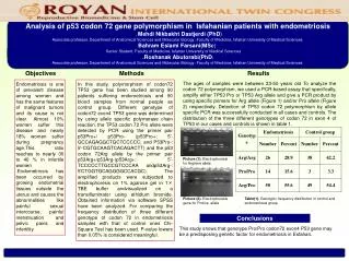
Immunohistochemical Analysis of p53 Tumor Suppressor Gene in Retinoblastoma Supriyo Ghose, Sumita Sethi, Parul Saxena, Seema Kashyap, Jasbir Kaur, Neelam Pushker Dr.R.P.Centre for Ophthalmic Sciences, All India Institute of Ophthalmic Sciences New Delhi E-poster (FP835) for The 69th Annual Conference of the All India Ophthalmological Society, Ahmedabad, Feb 3-6, 2011.
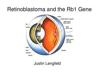
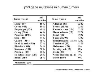
Introduction • Retinoblastoma is the most common primary intraocular malignancy in children. • A thorough understanding of the pathogenesis is important for its prevention and treatment. • Knudson’s ‘two-hit hypothesis’ initially laid the foundation for the understanding of genetics in retinoblastoma tumorigenesis.1 • Currently its accepted that the main genetic defect of retinoblastoma is associated with the RB1 gene.
Expression of p53 gene, a tumor suppressor gene located on chromosome 17 has been linked with the function of RBI gene. • Studies on animal models have shown that loss of both the Rb gene and p53 reactivity is required for development of malignancy.2 • We studied immunohistochemical expression of p53 tumor suppressor gene in retinoblastoma. 1. Knudson AG Jr. Mutation and cancer: statistical study of retinoblastoma. Proc Natl Acad Sci U S A. 1971 Apr;68(4):820-3. 2. Schlamp CL, Poulsen GL, Nork TM, Nickells RW. Nuclear exclusion of wild-type p53 in immortalized human retinoblastoma cells. J Natl Cancer Inst. 1997 Oct 15;89(20):1530-6.
Materials and methods • Eyes enucleated over a one year period from May 2008 to May 2009 as primary treatment for advanced intraocular retinoblastoma were included in the study. • Formalin fixed paraffin embedded sections were subjected to hematoxylin and eosin staining for studying histopathological features (tumor differentiation and high risk features). • p53 immunostaining was performed using mouse monoclonal antibody, in a working dilution of 1: 50. • p53 immunostaining graded semiquantitatively as negative, low or high expression.
Results Demographic details • Total 24 eyes • Age- Mean:2.45 Standard deviation: 1.3 Range 0.8 years to 6.5 years • Sex- Male:19 Female: 5 • Laterality- Unilateral:10 Bilateral: 14
Results Histopathological features: • Differentiation: Well differentiated: 9 Poorly differentiated: 15 • High risk histopathological features: Optic nerve beyond retrolaminar space: 8 Resected margin of the optic nerve: 7 Massive choroidal invasion: 11 Scleral invasion: 7 Iris involvement: 3 Anterior segment involvement: 5 Ciliary body involvement: 2 Extraocular involvement: 4
Results P53 immunostaining: • p53 immunostaining positive in 13/24 (54.1%) cases. • More than half of cases (7/13) with positive p53 immunostaining had strong positivity(53.8%). • No correlation of p53 expression with laterality, tumor differentiation and presence of high risk histopathological features.
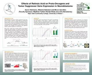
(a) (b) (c) (d) (a) Poorly differentiated retinoblastoma and (b) Well differentiated retinoblastoma showing numerous Flexner Wintersteiner rosettes (H and E staining). Cells showing nuclear positivity for p53 immunohistochemical staining (c) strong and (d)weakly positive.
Conclusion • p53 expressed in more than half (54.1%) of total retinoblastoma cases studied. • p53 strong expression in more than half of total p53 positive cases (53.8%). • p53 expression may not be associated with the development of primary retinoblastoma, but may play a role in progression of Retinoblastoma lesions. • p53 expression may thus be an indicator of aggressive tumor behavior.
More number of prospective studies with a long term follow up of children with high positive p53 expressions will help in understanding clinical significance of this finding.
Suggested reading • Schwimer CJ, Prayson RA. Clinicopathologic study of retinoblastoma including MIB-1, p53, and CD99 immunohistochemistry. Ann Diagn Pathol. 2001Jun;5(3):148-54. • Nigro JM, Baker SJ, Preisinger AC et al. Mutations in the p53 gene occur in diverse human tumour types. Nature. 1989 Dec 7;342(6250):705-8. • Simmons ML, Lamborn KR, Takahashi M et al. Analysis of complex relationships between age, p53, epidermal growth factor receptor, and survival in glioblastoma patients. Cancer Res. 2001 Feb 1;61(3):1122-8.