Download

1 / 46
510 likes | 1k Views
FIRST TRIMESTER BLEEDING. WALID SAGHIR M.D. FACS FICS Beirut, LEBANON Antalya May 15, 2014. Vaginal bleeding in pregnancy. 20-40% of all first trimester pregnancies About one half of those who bleed will miscarry. Etiology often not determined May be “normal” i.e. implantation bleeding

E N D
FIRST TRIMESTER BLEEDING WALID SAGHIR M.D. FACS FICS Beirut, LEBANON Antalya May 15, 2014
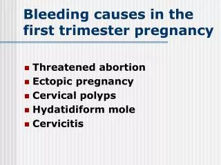
Vaginal bleeding in pregnancy • 20-40% of all first trimester pregnancies • About one half of those who bleed will miscarry. • Etiology often not determined • May be “normal” i.e. implantation bleeding • May be unrelated to pregnancy (cervicitis) • Goal: exclude pathology • Ectopic • Molar pregnancy
“Implantation bleeding” • Occurs 5-12 days after conception (prior to positive HCG test) • Lighter and shorter than normal period • Often mistaken for a “period” • Unrelated to pregnancy outcomes
Normal First Trimester Pregnancy Markers • Elevated B-hCG level produced by the placenta after implantation of the blastocyst. Occurs at ~ 23 menstrual days’ gestation, or as early as 8 days after conception. • Laboratory and transvaginalSonographic discriminatory findings
Yolk sac (YS) within the gestational sac at five to six menstrual weeks. This is the first sonographic finding that positively confirms intrauterine pregnancy
The embryo is first visible as a fetal pole adjacent to the yolk sac (YS). Cardiac activity is often visible at this time.
Measurement of the embryonic crown-rump length is the most accurate way to date pregnancy. This 10.5-week pregnancy measures 38 mm.
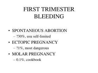
FIRST TRIMESTER BLEEDING • SPONTANEOUS ABORTION • ECTOPIC PREGNANCY • MOLAR PREGNANCY
Terminology of Abortion • Spontaneous abortion • Threatened abortion • Inevitable abortion • Complete abortion • Incomplete/missed abortion • Blighted ovum (anembryonic pregnancy) • Habitual/recurrent abortion
Definitions of abortion • Threatened Ab: Bleeding or cramping with closed cx in first half of pregnancy • Inevitable Ab: above + dilatation of cx • Complete Ab: all products have been expelled • Incomplete Ab (missed): POC remain in uterus • Septic ab: uterine infection with POC in uterus • Anembryonic pg: blighted ovum • Habitual abortion: 3 or more Ab’s in succession
SPONTANEOUS ABORTION • Incidence:~ 20% each pregnancy • Pregnancy loss prior to 20 weeks (500g) • PRIOR TO 6 WEEKS • Most likely chromosomal abnormality • AFTER 8 WEEKS - More likely due to maternal factors • Prognosis is good after, with 60% of patients having a viable pg next time
Risk Factors – maternally related • Age (50% rate > 45 y.o.) • Structural anomalies • Maternal infections • Endocrine problems • Autoimmune / coag problems • Blood group incompatibility • Severe malnutrition • Toxins (lead, smoking, ETOH, caffeine>300 mg/d, radiation, anesthesia)
Risk factors – pregnancy related • Gestational age • Subchorionic hemorrhage • Gestational sac size • Yolk sac • Fetal cardiac rate
HISTORY • LMP • AMOUNT & QUALITY OF FLOW • DATE OF POSITIVE HCG • PAIN? • PREVIOUS ABORTIONS
DIAGNOSIS • Pelvic exam • Cx – dilated? • Amount of bleeding • Uterine size • Pain with palpation • Adnexal masses • LABS • sHCG – quantitative • Rh & type • Hemogram • Progesterone (?) • Ultrasound
QUANTITATIVE HCG • Serial measurements - Doubles q 48-72 hrs (>66% in 48 hours) - doubling? Plateau? Falling? • Correlation with dates - LMP/first positive Pg test/uterine size • Correlation with US findings
HCG levels • Gestational sac: 1000 – 1500 • Yolk sac : 5000 • Cardiac activity: 10,000 (?) – 15,000
ULTRASOUND • Gestational sac – IUP? • Yolk sac – normal or abnormal? • Embryo – know what to tell patient - Embryo may not be expected - If FHB (+) is seen – 90-96% go to term • Subchorionic bleeding
Management of the patient • Bed rest does not prevent loss • Pelvic rest is advised • Even though nothing can be done, constant f/u with patient is warranted – mainly for psychological reasons • Anticipate complications/heavy bleeding • If Rh negative give Rhogam within 72 hours • D & C
Recurrent Abortion • 3 more consecutive pregnancy losses<20 weeks • ~1% of patients • Most likely due to - Chromosomal factors - Uterine abnormalities - SLE or other autoimmune problems - Thyroid disease
ECTOPIC PREGNANCY • 1/250 – 1/87 pregnancy • 15% of all maternal mortality • 98% occur in the fallopian tube • 2% other portions of cornua, cx, ovary, abd • Increased incidence with risk factors
RISK FACTORS FOR ECTOPIC PREGNANCY • Risk factor
RISK FACTORS FOR ECTOPIC PREGNANCY Risk factor Odds Ratio • Previous ectopic • Tubal surgery • BTL • IUD • Infertility • DES exposure • Previous STD • Previous abd surgery • - 9.3 – 47 • - 6.0 – 11.5 • - 3.0 – 139 • - 4.2 – 45 • - 1.1 – 28 • - 2.4 – 13 • - 2.5 – 3.7 • - 0.9 – 3.8
Clinical presentation • Pelvic/abd pain – 100% • Tenderness on exam – 80% • Bleeding – 75% • Amenorrhea – 74% • Adnexal mass – 50%
DIAGNOSIS • AWARENESS OF THE POSSIBILITY • History • Pelvic exam • S-HCG • Ultrasound
History • High index of suspicion • Same as for threatened abortion • ALWAYS ask Q’s related to risk • ALWAYS ask about pelvic pain
Physical Examination • Orthostatic changes • Pelvic exam - Document . Abdominal pain . Adnexal masses/pain . Cervical motion tenderness . Uterine size - IF EARLY, exam may be NORMAL
DIAGNOSIS • HCG > 1500 with no IUP • Pos. HCG/pelvic pain/vag bleeding 5-7 weeks pg • HCG not doubling in pg with vaginal bleeding • Potential in any pt with pos. HCG and pelvic pain
DIAGNOSIS • Ultrasound - Evidence of DEFINITIVE intrauterine GS excludes ectopic - Pseudosac - Empty uterus – get HCG for correlation . May need to repeat the history/PE daily . Serial HCG’s and US
HETEROTOPIC PREGNANCY • RARE – 1:30,000 ECTOPIC PREGNANCIES
TREATMENT AND CRITERIA FOR MANAGING ECTOPIC PREGNANCY Expectant management • No evidence of tubal rupture • Minimal pain or bleeding • Patient reliable for follow-up • Starting β-hCG level less than 1,000 mIU per mL (1,000 IU per L) and falling • Ectopic or adnexal mass less than 3 cm or not detected • No embryonic heartbeat
Medical management with methotrexate • Stable vital signs and few symptoms • No medical contraindication for methotrexate therapy (e.g., normal liver enzymes, complete blood count and platelet count) • Unruptured ectopic pregnancy • Absence of embryonic cardiac activity • Ectopic mass of 3.5 cm or less • Starting β-hCG levels less than 5,000 mIU per mL (5,000 IU per L) • Dosage: single intramuscular dose of 1 mg per kg, or 50 mg per m2 • Follow-up: β-hCG on the fourth and seventh posttreatment days, then weekly until undetectable, which usually takes several weeks • Expected β-hCG changes: initial slight increase, then 15 percent decrease between days 4 and 7; if not, repeat dosage or move to surgery • Special consideration: prompt availability of surgery if patient does not respond to treatment
Combination gefitinib and methotrexate compared with methotrexate alone to treat ectopic pregnancy. Skubisz MM1, Horne AW, Johns TG, Nilsson UW, Duncan WC, Wallace EM, Critchley HO, Tong S. Author information Abstract OBJECTIVE: To determine the safety, tolerability, and efficacy of combination gefitinib and methotrexate to treat ectopic pregnancy. METHODS: We performed a phase I, single-arm (nonrandomized), open-label study. Twelve women with ectopic pregnancies were administered methotrexate (50 mg/m, intramuscular) and 250 mg oral gefitinib in a dose-escalation protocol: one dose (day 1) n=3; three doses (days 1-3) n=3; seven doses (days 1-7) n=6. Efficacy was examined by comparing human chorionic gonadotrophin (hCG) decline and time to resolution with historic controls administered methotrexate only. RESULTS: Common side effects were transient acneiform rash in 67% (8/12) and diarrhea in 42% (5/12) of participants. There was no clinical or biochemical evidence of serious pulmonary, renal, hepatic, or hematologic toxicity. Of six participants with a pretreatment serum hCG level between 1,000 and 3,000 international units/L, hCG levels declined significantly faster than in the control group. Median serum hCG levels by day 7 after treatment were less than one fifth of levels observed among 71 historic controls treated with methotrexate alone (median [interquartile range] hCG in participants 261 [55-1,445] international units/L compared with controls 1,426 [940-2,573]; P=.008). Median time for the ectopic pregnancies to resolve with combination therapy was 34% shorter compared with methotrexate alone (21 days compared with 32 days; P=.018). CONCLUSION: Combination gefitinib and methotrexate has potential as a treatment for ectopic pregnancy but is commonly associated with minor side effects such as transient rash and diarrhea. The treatment requires validation of safety and efficacy in a larger trial. CLINICAL TRIAL REGISTRATION: Australian New Zealand Clinical Trials Registry, www.anzctr.org, AC'TRN12610000684022. LEVEL OF EVIDENCE: : II.
Surgical management • Unstable vital signs or signs of hemoperitoneum • Uncertain diagnosis • Advanced ectopic pregnancy (e.g., high β-hCG levels, large mass, cardiac activity) • Patient unreliable for follow-up • Contraindications to observation or methotrexate
HYDATIFORM MOLE or Gestational trophoblastic disease (GTD) • 1:1500 – 1:2000 pregnancies • More common in pts>40 y.o. • Complete vs. partial • Historically diagnosed later • Potential for metastases
DIAGNOSIS • sHCG high (hyperemesis common) • Uterus large for dates • Vaginal bleeding • Ultrasound – typical US presentation • “Grape clusters”
TREATMENT • Surgical evacuation • Tissue to pathology • Serial HCG’s until negative X 1 yr • Pregnancy contraindicated X 1 yr • No contraindication to OCP use • Very slight increased risk of mole again • CXR