Download
1 / 24
310 likes | 863 Views
SHIGELLA & PROTEUS. By. Dr. Emad AbdElhameed Morad. Lecturer of Medical Microbiology and Immunology. SHIGELLA. Morphology: Gram negative bacilli, non motile, non capsulated. Cultural characters: On MacConkey’s medium: pale non lactose fermenting colonies.

E N D
SHIGELLA & PROTEUS By Dr. Emad AbdElhameed Morad Lecturer of Medical Microbiology and Immunology
SHIGELLA Dr. Emad AbdElhameed Morad
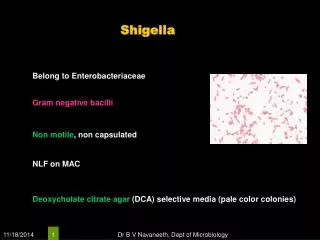
Morphology: • Gram negative bacilli, non motile, non capsulated. • Cultural characters: • On MacConkey’s medium: pale non lactose fermenting colonies. • On desoxycholate citrate agar medium: similar to MacConkey’s medium. Dr. Emad AbdElhameed Morad
Biochemical reactions: • Sh. dysenteriae ferments glucose only with production of acid only. • Other shigella species ferment glucose and mannite with acid production only. • Shigella sonnei is a late lactose fermenter. • All shigella are H2S negative and urease negative. • Serological characters: • They are divided into four groups according to O antigen: • Group A: Sh. dysenteriae includes 13 serotypes. • Group B: Sh. flexneri includes 8 serotypes. • Group C: Sh. bodyii includes 18 serotypes. • Group D: Sh. sonnei includes one serotype. Dr. Emad AbdElhameed Morad
Virulence factors: • Invasiveness of the mucosa of distal ileum and colon. Note that shigella does not invade the blood stream. • Shiga toxin produced by Shigella dysenteriae type I: • It is heat labile exotoxin that affects both gut and CNS. • It acts as an enterotoxin causing diarrhea similar to E. coli verotoxin. • It acts also as a neurotoxin causing meningism and coma in severe fatal infections. Dr. Emad AbdElhameed Morad
Diseases caused by shigella: • Shigella causes shigellosis or bacillary dysentery. • It is only a human disease. • No animal reservoir. • The organism is transmitted by : 4 F Fingers Faeces Flies Food Dr. Emad AbdElhameed Morad
The incubation period is 1 – 4 days. • The shiga toxin acts on gut to produce early non bloody diarrhea. • Then, invasion of the mucosa occurs leading to late dysentery which means diarrhea + mucous + blood + pus. • Symptoms are:fever, abdominal pain, diarrhea (early non bloody, late dysentery) and tenesmus. • The stool contains blood, mucous, pus. • Recovery occurs spontaneously in most cases. • However, in children and elderly, dehydration, acidosis and death may occur. • Very few cases remain as chronic carriers. Dr. Emad AbdElhameed Morad
Diagnosis of shigella infections: • Specimen: • Stool. • Macroscopic examination: • Contains mucous, blood. • Microscopic examination: • Pus cells and red blood cells. • Culture on the following media: • MacConkey’s agar or DCA • Selenite broth for 24 hours after which subculture is done on MacConkey’s medium or DCA. • Identification of the growing colonies is done by: Dr. Emad AbdElhameed Morad
Colonial morphology Pale colonies Gram stain Gram negative bacilli Shigella dysenteriae ferments glucose with acid production only Biochemical reactions Other shigella species ferment glucose and mannite with acid production only H2S negative, urease negative and non motile Serological identification Latex agglutination test with specific antisera Dr. Emad AbdElhameed Morad
Prevention of shigella infections: • Proper sewage disposal. • Chlorination of water. • Good personal hygiene. • Treatment of shigella infections: • Fluid and electrolyte balance. • Ciprofloxacin is the drug of choice. • But, antibiotic susceptibility should be done because drug resistance is very high among shigella strains. Dr. Emad AbdElhameed Morad
Proteus Dr. Emad AbdElhameed Morad
Morphology: • Gram negative bacilli, highly motile, very pleomorphic. • Cultural characters: • On nutrient agar:colonies with swarming growth in successive waves + Fishyodor. • On MacConkey’s agar:Pale non lactose fermenting colonies. The swarming is inhibited on this medium. Swarming could also be inhibited on blood agar containing phenyl ethyl alcohol. Dr. Emad AbdElhameed Morad
(Proteus on nutrient agar) Dr. Emad AbdElhameed Morad
(Proteus on nutrient agar) Dr. Emad AbdElhameed Morad
Biochemical reactions: • Phenylalanine deaminase +Ve. • Urease +Ve. • H2S +Ve. • Indole +Ve except Pr. mirabilis. • Serological characters: • Certain types of proteus (OX19, OXK, OX2) have antigens in common with rickettsiae. So, suspensions of these strains are used in diagnosis of rickettsial infections in Weil-Felix reaction. However, this test is non specific. Dr. Emad AbdElhameed Morad
(Phenylalanine deaminase test) +Ve -Ve Dr. Emad AbdElhameed Morad
(Urease test) +Ve -Ve Dr. Emad AbdElhameed Morad
Species of proteus: • Proteus vulgaris. • Proteus mirabilis. • These species are found as normal flora in the colon and also in soil and water. • These species cause the following diseases: • Urinary tract infections (particularly Proteus mirabilis). • Wound infections. • Otitis media. • Pneumonia. • Meningitis. • Bacteremia. • Infections may be hospital or community acquired. Dr. Emad AbdElhameed Morad
Providencia rettgeri Morganella morganii • What about ? • These bacteria were previously included under proteus as Proteus morganii and Proteus rettgeri because they are similar to proteus in being Gram negative bacilli, non-lactose fermenters, phenylalanine deaminase positive, urease positive and indole positive. But they are H2S negative. • Recently they have been separated from proteus into separate genera based on molecular DNA studies. • They are resistant to antibiotics and now emerging as important nosocomial pathogens. Dr. Emad AbdElhameed Morad
Diagnosis of proteus infections: • Specimen: • Urine, pus, CSF, etc. • Direct smear: • Direct smear stained with Gram stain shows Gram negative bacilli among pus cells. • Culture on nutrient agar • Swarming growth + fishy odor. • Culture on MacConkey’s agar • Identification of the growing colonies is done by: Dr. Emad AbdElhameed Morad
Colonial morphology Pale non lactose fermenting colonies Gram stain Gram negative bacilli Phenylalanine deaminase +Ve, urease +Ve, H2S +Ve, indole +Ve except Proteus mirabilis Biochemical reactions Motility test Highly motile bacteria Dr. Emad AbdElhameed Morad
Treatment of proteus infections: • Proteus infections are resistant to antibiotics and so antibiotics should be given according to the results of antibiotic sensitivity tests. • However, most strains are sensitive to aminoglycosides and trimethoprim-sulfamethoxazole. Dr. Emad AbdElhameed Morad
Acinetobacter • Acinetobacter is oxidase negative, non fermentative, non motile Gram negative cocco bacilli found in soil and water and also can be part of normal flora. • Species of acinetobacter are Acinetobacter calcoaceticus and Acinetobacter baumannii. • They are opportunistic pathogens that cause infections in hospitalized patients especially those immunocompromised with catheters. Dr. Emad AbdElhameed Morad
Thank you Dr. Emad AbdElhameed Morad