Download
1 / 14
140 likes | 303 Views
Application of Mismatch Detection Methods in DNA Computing. C. Henkel et al. Leiden University, The Netherlands. Introduction. One focus of biomolecular computing studies is the use of combinatorial libraries of DNA to provide search spaces for parallel filtering algorithms.

E N D
Application of Mismatch Detection Methods in DNA Computing C. Henkel et al. Leiden University, The Netherlands
Introduction • One focus of biomolecular computing studies is the use of combinatorial libraries of DNA to provide search spaces for parallel filtering algorithms. • For example, The blocking algorithm uses DNA complementarity to remove molecules not representing a solution for the formal satisfiability problem from the candidate pool. • The original proposal for blocking algorithm was using PCR inhibition through association with blocker molecules, for example PNA.
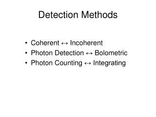
Heteroduplex Migration Assay • Homoduplex • perfect double stranded DNA • during electrophoresis, migrates through a gel at a predictable rate • Heteroduplex • DNA containing nucleotide mismatches • migrates at an anomalous rate, caused by secondary structure formation or helix distortion. • mobility of heteroduplexes is lower than that of homoduplexes of equal length. • Several well-established and sensitive mutation detection techniques exploit this effect. • single strand conformational polymorphism (SSCP), temperature or denatureing gradient gel electrophoresis (TGGE,DGGE) and heteroduplex analysis
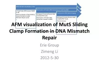
Enzymatic Mismatch Recognition • also widely used in mutation detection. • uses specific endonucleases which recognize and digest the abnormal DNA conformations which result from mismatched nucleotides. • Here, used the recently discovered CEL I nuclease, purified from celery [Yang,B. et al., Biochemistry (2000)]
Problem Instance and Algorithm • tested mutation detection techniques on the following 4 variable, 4 clause satisfiability (3-CNF-SAT) problem. F=(¬a∨b∨¬c)∧(a∨¬b∨d)∧(¬a∨c∨¬d)∧(b∨c∨¬d) • The blocking algorithm • synthesize all possible assignments as ssDNAs • synthesize blockers representing to falsifying assignments • mix and hybridize • apply mismatch detection method
Sequence Design • Assignments • 5’-[S][a][b][c][d][F]-3’ • S=GAACGA, true=GTCTGA, false=TAGTGG, F=AACGTG • Blockers • 3’-[S][a][b][c][d][F]-5’ • ¬a∨b∨¬c 1010,1011 A0,A1 • a∨¬b∨d 0100,0110 B0,B1 • ¬a∨c∨¬d 1001,1101 C0,C1 • b∨c∨¬d 0001,1001 identical to C0
Oligonucleotides • Oligonucleotides were custom synthesized and labeled at Isogen Bioscience. • For detection, library molecules contain a covalent 5’ Cy5 label (Amersham Biosciences), blockers a 5’ fluorescein (FIFC, Molecular Probes). • All oligos were purified from 10% denaturing polyacrylamide gels to remove unbound dyes. • DNA was allowed to diffuse from gel slices by overnight soaking. • Concentrations were calculated from absorption measurements of the dyes at 494 nm (fluorescein) and 649 nm (Cy5) • Molar extinction coefficients of 77,000 cm-1M-1 (fluorescein) and 250,000 cm-1M-1 (Cy5) were used.
Duplex Migration Assay • Mixtures of library and blocker molecules were made by combining 5 pmol per oligo in a gel loading buffer consisting of 1x TBE, 3.3% sucrose and 0.033% Orange G. • Duplex DNA was formed by heating the mixtures to 95℃ for 5 minutes, and cooling to 4 ℃ at 0.1℃/sec in a thermocycler (Biometra Tgradient). • Gels were prepared from regular acrylamide: bisacrylamide (20:1) or proprietary SequaGel MD (Mutation Detection) acrylamide matrix (National Diagnostics, Atlanta, Georgia, USA). • Gels were run in 1x TBE at 200V and 4℃. Gel images were captures on a a Biorad FluorS MultiImager, using UV excitation with 530 nm band pass and 610 nm long pass filters for detection of fluorescein and Cy5 fluorescence, respectively. • Contrast levels of digital images were adjusted in Corel Photopaint 11.
Enzymatic Mismatch Cleavage Assay • Duplexes were prepared as above, except that hybridization was carried out in 10 mM Tris/HCl pH 8.5. • T7 endonuclease I (T7E1) was obtained from New England Biolabs and handled according to the manufacturers recommendations. Reactions containing 5 pmol per oligo and 1 unit of enzyme were allowed to proceed for up to 150 minutes. • CEL I enzyme was obtained from DR. Edwin Cuppen (Hubrecht Laboratory, Utrecht, NL; http://cuppen.niob.knaw.nl). • Every lot of CEL I was tested, and for all subsequent experiments quantities were used that gave the effect shown in Figure 3 after 30 minutes of incubation. Reaction were performed with 5 pmol per oligo in a 4 μl volume at 45℃, in a 10 mM MgSO4, 10 mM HEPES pH 7.5, 10 mM KCl, 0.002% Triton χ-100, 0.2 μg/μl BSA buffer. Reactions were stopped by placing samples on ice and adding 4 μl 80% formamide, 100 mM EDTA. • Digests were analyzed on 10% TBE/polyacrylamide gels, which were imaged as before. Bands were anlyzed using ImageJ software
Result-heteroduplex migration • 12.5% acrylamide gels supplemented with 20% urea were found to give good separation. • Arrows indicates apparent homoduplexes. • Every blocker should only be able to form a perfect duplex with one of the library oligos, but… • No improvement was found using MD gel matrix or longer gels • Nonetheless, oligo 0,2,8 don’t behave as a homoduplex.
Result-mismatch endonucleases • T7EI did not have any discernable effect. (here, 0.2 units were used per reaction; 1 unit per reaction gave identical results) • CEL I has a clear effect on all samples. • It completely degrades the mismatched DNA within 30 minutes. • Perfect dsDNA is still detectable after 150 minutes
Discussion • Differential duplex migration did not provide a suitable test system. But, there is no general theory describing the effect of anomalous DNA conformations on migration rate, and it was already known that not all mismatches can be detected this way. • With careful optimization of the encoding, the use of longer molecules and more sophisticated analytical techniques (e.g. capillary electrophoresis), the method holds considerable promise.
Future work • Several other proteins that bind mismatches (such as MutS) do not destroy the DNA molecule. Such proteins may be used in a gel-shift assay. • Besides the enzymatic method tested here, chemical cleavage of mismatches could be considered.