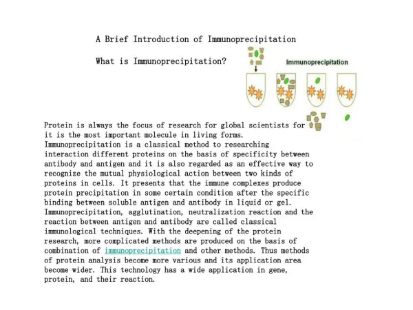
Download
1 / 3
30 likes | 143 Views
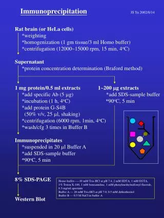
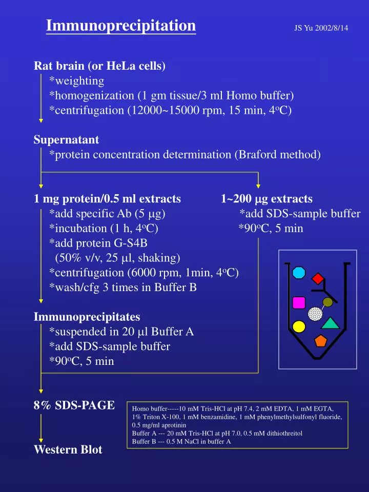
Immunoprecipitation JS Yu 2002/8/14. Rat brain (or HeLa cells) *weighting *homogenization (1 gm tissue/3 ml Homo buffer) *centrifugation (12000~15000 rpm, 15 min, 4 o C) Supernatant *protein concentration determination (Braford method)

E N D
ImmunoprecipitationJS Yu 2002/8/14 Rat brain (or HeLa cells) *weighting *homogenization (1 gm tissue/3 ml Homo buffer) *centrifugation (12000~15000 rpm, 15 min, 4oC) Supernatant *protein concentration determination (Braford method) 1 mg protein/0.5 ml extracts1~200 mg extracts *add specific Ab (5 mg) *add SDS-sample buffer *incubation (1 h, 4oC) *90oC, 5 min *add protein G-S4B (50% v/v, 25 ml, shaking) *centrifugation (6000 rpm, 1min, 4oC) *wash/cfg 3 times in Buffer B Immunoprecipitates *suspended in 20 ml Buffer A *add SDS-sample buffer *90oC, 5 min 8% SDS-PAGE Western Blot Homo buffer-----10 mM Tris-HCl at pH 7.4, 2 mM EDTA, 1 mM EGTA, 1% Triton X-100, 1 mM benzamidine, 1 mM phenylmethylsulfonyl fluoride, 0.5 mg/ml aprotinin Buffer A --- 20 mM Tris-HCl at pH 7.0, 0.5 mM dithiothreitol Buffer B --- 0.5 M NaCl in buffer A
PAK2 (N17) Antibody Background: p21-activated kinases (PAKs) were initially characterized by Manser et al. (1) as a set of 62-68 kDa proteins with an unique property that they can bind to small (21 kDa) guanosine triphosphatases (GTPases) Rac and Cdc42 that regulate actin polymerization. At least three isoforms of PAK (PAK1~3) have been identified in mammalian tissues (2). After binding to active form of Rac or Cdc42, PAKs undergo autophosphorylation/activation process and become as active kinases capable of acting on exogenous substrates (1). Another regulation mechanism of PAKs involves proteolytic removal of its N-terminal regulatory region (3, 4). Recent studies have indicated that PAKs are involved in modulating diverse cell functions, including cytoskeleton rearrangement, apoptotic cell death and cell cycle progression. In addition, PAKs can be activated in cells by various extracellular stimuli such as growth factors, chemoattractant, thrombin, angiotensin II and CD28, and can act as upstream regulators of the MAPK, JNK and p38 MAPK pathways. These observations suggest that PAKs are important enzymes that participate in multiple cellular signaling pathways. Specificity/Sensitivity: PAK2 (N17) antibody detects total endogenous levels of PAK2 (62 kDa). The antibody does not cross-react with other PAK isoforms. Source/Purification: Polyclonal antibodies are produced in rabbits by using the peptide, MSDNGELEDKPPAPPVR, corresponding to the NH2-terminal region from amino acids 1-17 of the sequence of human and rabbit PAK2 as the antigen. A cysteine residue was added to the C-terminus to facilitate coupling the peptide to Keyhole Limpets hemocyanin (KLH) or bovine serum albumin (BSA). Antibodies are purified by peptide affinity chromatography. Applications: PAK2 (N17) antibody can be used in Western blot and immunoprecipitation. (A) (B) ppt sup - + - + (N17 peptide) A431 Hep3B Balb/3T3 PAK2 PAK2 (A) Western blot analysis of extracts from human A431, Hep3B and Blab/3T3 cells. (B) Immunoprecipitation of PAK2 from A431 cells extracts in the absence (-) and presence (+) of N17 peptide, followed by western blot analysis using PAK2 (N17) antibody. References:(1) Manser, E. Et al. (1994) Nature 367, 40-46. (2) Sells, M. A. and Chernoff, J. (1997) Trends Cell Biol. 7, 162-167. (3) Rudel, T. and Bokoch, G. M. (1997) Science276, 1571-1574. (4) Chan, W.-H., and Yu, J.-S. and Yang, S.-D. (2000) Biochem. J.351, 221-232.
Anti-peptide Ab of human PAK2 (62 kDa) 1-17 IP/WB using A431 cell extracts ppt sup ppt - + - + peptide + - B E E 2nd Antibody-AP Biotin-strepavidin-AP