Download
1 / 104
1.04k likes | 1.22k Views
Dissection of the Anterior Compartment of the Forearm.

E N D
Place the cadaver in the supine position. Make a vertical skin incision down the center of the anterior surface of the forearm from the cubital fossa to the distal transverse crease of the wrist. Incise the skin transversely across the front of the wrist. Reflect the skin flaps medially and laterally. Identify the cephalic and basilic veins and the lateral and medial cutaneous nerves of the forearm. Remove the superficial fascia along the lines of the skin incision.
Cut the bicipital aponeurosis and remove the deep fascia of the anterior compartment of the forearm along the lines of the skin incisions. Do not damage the underlying structures.
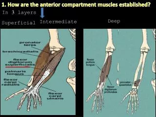
Identify again the brachiordialis muscle and confirm that it arises from the upper two-thirds of the lateral supracondylar ridge of the humerus. This muscle is situated in the lateral fascial compartment of the forearm and will be considered later.
Identify the following superficial group of muscles in the anterior fascial compartment and note that they possess a common tendon of origin which is attached to the medial epicondyle of the humerus. The muscles are named as follows, from lateral to medial:
1. Pronator teres, with its humeral and ulnar heads. 2. Flexor carpi radialis. 3. Palmaris longus (not always present). 4. Flexor carpi ulnaris.
Now transect the humeral head of the pronator teres as it crosses the underlying median nerve. At the same level, transect the bellies of the flexor carpi radialis and the palmaris longus muscles. The flexor digitorum superficialis muscle is now exposed.
Note that the pronator teres is supplied by a branch of the median nerve that arises in the cubital fossa. The flexor carpi radialis and the palmaris longus are supplied by branches of the median nerve deep to the pronator teres. The flexor carpi ulnaris is supplied by branches of the ulnar nerve as it enters the forearm by passing behind the medial epicondyly of the humerus.
Intermediate Flexor Muscle. Study now the flexor digitorum superficialis muscle. Note that the humeroulnar head arises from the common tendon attached to the medial epicondyle of the humerus and the medial margin of the coronoid process of the ulna; it is also attached to the medial ligament of the elbow joint. A radial head arises from the oblique line on the anterior surface of the shaft of the radius. Observe that the two heads are connected by a fibrous arch beneath which pass the median nerve and the ulnar artery.
Deep Flexor Muscles. Transect the distal portion of the belly of the flexor digitorum superficialis just proximal to the tendons. Avoid cutting the median nerve, which is bound to the deep surface of the muscle with fascia. Reflect the muscle and thus expose the deep muscles in this compartment, namely, the flexor pollicis longus, flexor digitorum profundus, and pronator quadratus muscles.
Now expose and clean the main arteries in the anterior compartment.
The radial artery, one of the terminal branches of the brachial artery, arises in the cubial fossa and passes downward and laterally overlapped by the brachioradialis muscle. Observe that in the lower half of the forearm the radial artery emerges on the medial side of the brachioradialis tendon and then lies on the lateral side of the tendon of the flexor carpi radialis.
Here, the radial artery is covered only by skin and fascia, and it rests posteriorly on the anterior surface of the distal part of the radius. Note that in the middle third of its course the superficial branch of the radial nerve lies lateral to it. The radial artery leaves the forearm by winding around the lateral aspect of the wrist to reach the posterior surface of the hand.
1.Muscular branches to neighnoring muscles. 2. Recurrent branch, which joins the arterial anastomosis around the elbow joint. 3. Superficial palmar branch, which arises above the wrist and enters the palm of the hand.
The ulnar artery, one of the terminal branches of the brachial artery, arises in the cubital fossa and passes downward, deep to the superficial flexor muscles and the flexor digitorum superficialis.
At the wrist it emerges between the tendon of the flexor carpi ulnaris and the tendon of the flexor digitorum superficialis muscle. Note that in the lower two-thirds of its course the ulnar nerve lies on the medial side of the flexor digitorum superficialis muscle. The ulnar artery enters the palm on the lateral side of the pisiform bone.
1.Muscilar branches to neighboring muscles. 2. Recurrent branches that join the arterial anastomosis around the elbow joint. 3. Anastomotic branches that take part in the arterial anastomosis around the wrist joint.
4. The common interosseous artery, which arises from the upper part of the ulnar artery and quickly divides into the anterior and posterior interosseous arteries. The anterior interosseous artery is small in size and should be followed down the anterior surface of the interosseous membrane between the flexor pollicis longus and the flexor digitorum profundus.
Median nerve.Now trace the course of the median nerve in the forearm. The median nerve leaves the cubital fossa by passing between the two heads of pronator teres. It continues downward adherent to the posterior surface of the flexor digitorum superficialis. At the wrist it emerges between the lateral margin of the tendons of flexor digitorum superficialis and the tendon of flexor carpi radialis. Here it lies posterior to the palmaris longus tendon. The median nerve enters the palm by passing posterior to the flexor retinaculum.
1. Muscular branches in the cubital fossa to the pronator teres, the flexor carpi radialis, the palmaris longus, and the flexor digitorum superficialis.
3. Anterior interosseous nerve, which arises from the median nerve as it emerges from between the two heads of the pronator teres muscle. It runs down on the anterior surface of the interosseous membrane. Trace branches from it to the flexor pollicis longus, the lateral half of flexor digitorum profundus, and the pronator quadratus.
4. Palmar cutaneous branch, Which passes to the skin over the lateral part of the palm.
Ulnar nerve.The ulnar nerve enters the forearm from behin the medial epicondyle of the humerus. Note that it crosses the medial ligament of the elbow joint and passes between the two heads of the flexor carpi ulnaris. Trace the nerve downward between the flexor carpi ulnaris and the flexor digitorum profundus muscles. At the wrist observe that the nerve lies between the tendons of the flexor carpi ulnar and the flexor digitorum superficialis muscles. The ulnar nerve enters the palm lateral to the pisiform bone, anterior to the flexor retinaculum.
1. Muscular branches to the flexor carpi ulnaris and to the medial half of the flexor digitorum profundus.
3. Palmar cutaneous branch, which arises in the middle of the forearm and supplies the skin over the hypothenar eminence.
4. Dorsal branch, or posterior cutaneous branch, which is large and passes medially between the tendon of flexor carpi ulnaris and the ulna and is distributed on the posterior surface of the hand and fingers.
Clean the brachioradialis muscle and verify that it arises from the upper two-thirds of the lateral supracondylar ridge of the humerus and from the adjoining lateral intermuscular septum. Note that its tendon is inserted into the base of the styloid process of the radius.
Identify and clean the extensor carpi radialis longus muscle and note that it arises from the lower third of the lateral supracondylar ridge of the humerus and from the adjoining laterl intermuscular septum. Its tendon of insertion will be examined later.
Radial Nerve. Review the course of the radial nerve from the point where it pierces the lateral intermuscular septum and passes forward into the cubital fossa. Note that at the level of the lateral epicondyle the radial nerve divides into superficial and deep branches.
Identify and clean the following branches of the radial nerve:
1. Muscular branches to the brachioradialis, to the extensor carpi radialis longus, and to the lateral part of the brachialis muscle.
3. Deep branches of the radial nerve. The deep branch passes between the superficial and deep layers of the supinator muscle and enters the forearm by winding round the neck of the radius.
4. Superficial branch of the radial nerve, which runs down the forearm beneath the brachioradialis muscle. In the lower part of the forearm, trace the nerve backward under the tendon of the brachioradialis. It supplies the skin on the dorsum of the hand and dorsum of the lateral two and one-half fingers.