Download
1 / 1
10 likes | 77 Views
High Resolution and High Efficiency Open SPECT Detector for Molecular Imaging Studies of Cardiovascular Diseases on Mice. 1.3 mm. 0.8 mm. 1.2 mm. 0.9 mm. 1.1 mm. 1.0 mm. Spine. Liver.

E N D
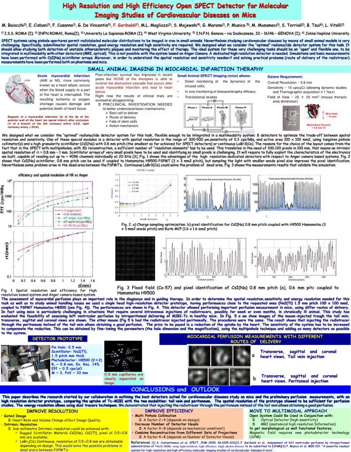
High Resolution and High Efficiency Open SPECT Detector forMolecular Imaging Studies of Cardiovascular Diseases on Mice 1.3 mm 0.8 mm 1.2 mm 0.9 mm 1.1 mm 1.0 mm Spine Liver M. Baiocchi1), E. Cisbani1), F. Cusanno1), G. De Vincentis3), F. Garibaldi2), M.L. Magliozzi1), S. Majewski4), G. Marano1), P. Musico 5), M. Musumeci1), S. Torrioli1), B. Tsui6), L. Vitelli1) • 1) I.S.S. ROMA (I); 2) INFN ROMA1, Roma(I), 3) University La Sapienza ROMA (I); 4) West Virginia University; 5) I.N.F.N. Genova – via Dodecaneso, 33 – 16146 – GENOVA (I); 6) Johns Hopkins University SPECT systems using pinhole apertures permit radiolabeled molecular distributions to be imaged in vivo in small animals. Nevertheless studying cardiovascular diseases by means of small animal models is very challenging. Specifically, submillimeter spatial resolution, good energy resolution and high sensitivity are required. We designed what we consider the “optimal” radionuclide detector system for this task. It should allow studying both detection of unstable atherosclerotic plaques and monitoring the effect of therapy. The ideal system for these very challenging tasks should be an “open” and flexible one, to be integrated in multimodality with other detectors (MRI, optical). This is not possible with the system based on Anger Camera. A dedicated high resolution detector is needed. Simulations and basic measurements have been performed with CsI(Na) scintillator arrays. Moreover, in order to understand the spatial resolution and sensitivity needso=f and solving practical probems (route of delivery of the radiotracer) measurements have been performed both on phantoms and mice. SMALL ANIMAL IMAGING IN MIOCARDIAL INFARCTION THERAPHY • Post-infarction survival has improved in recent years butNONE of the therapies is able to reverse the destructive cascade that occurs after acute myocardial infarction and lead to heart failure. • Right now the results of clinical trials are somewhat disappointing: • PRECLINICAL INVESTIGATION NEEDED to better understand basic mechanisms: • Best cell to deliver • Route of delivery • Fate of stem cells • Action mechanism Acute myocardial infarction(AMI or MI), more commonly known as a heart attack, occurs when the blood supply to a part of the heart is interrupted. The resulting ischemia or oxygen shortage causes damage and potential death of heart tissue. Small Animal SPECT Imaging (mice) allows: • Direct monitoring of the dynamics of the infused cells; • In vivo monitoring of disease/teraphy efficacy; • Translational studies Sistem Requirement: Overall Resolution ~ 0.8 mm Sensitivity ~ 10 cps/μCi (allowing dynamic studies and Thomographic acquisition ≤ 1 hour) Field of View ~ 25 × 25 mm2 (mouse thoracic area dimension) Diagram of a myocardial infarction (2) of the tip of the anterior wall of the heart (an apical infarct) after occlusion (1) of a branch of the left coronary artery (LCA, right coronary artery = RCA). We designed what we consider the “optimal” radionuclide detector system for this task, flexible enough to be integrated in a multimodality system: 8 detectors to optimize the trade-off between spatial resolution and sensitivity. One of these special modules is a detector with spatial resolution in the range of 300-500 μm,sensitivity of 0.3 cps/kBq, and active area 100 x 100 mm2, using tungsten pinhole collimator(s) and a high granularity scintillator (CsI(Na) with 0.8 mm pitch (the smallest so far achieved for SPECT detectors) or continuous LaBr3(Ce). The reasons for the choice of the layout comes from the fact that in the SPECT with multipinholes, with 3D reconstruction, a sufficient number of “resolution elements” has to be used. This translates in the need of 100-120 pixels in 100 mm, that means an intrinsic spatial resolution of ri = 0.8 mm – 1 mm. Scintillator arrays of very small pixels have to be used and identifying so small pixels is challenging. It will require to fully exploit the characteristics of the electronics we built, capable of reading out up to ~ 4096 channels individually at 20 KHz [3]. Fig. 1 shows the advantages of the high resolution dedicated detectors with respect to Anger camera based systems. Fig. 2 shows that CsI(Na) scintillator, 0.8 mm pitch can be used if coupled to Hamamatsu H9500 PSPMT (3 x 3 mm2 pitch), but sampling the light with smaller anode pixel size improves the pixel identification. Nevertheless some problem arise in the dead area between the PSPMT’s. Continuous LaBr3(Ce) could solve the problem of dead area. Fig. 3 shows the measurements results that validate the simulation. c b a Fig. 2. a) Chargesamplingoptimization. b) pixel identificationforCsI(Na) 0.8 mm pitchcoupledwith H9500 Hamamatsu (3x3 mm2 anodepitch) and Burle MCP (1.6 x 1.6 mm2 pitch) Fig. 3 Flood field (Co-57) and pixel identification of CsI(Na) 0.8 mm pitch (a), 0.6 mm pitc coupled to Hamamatsu H9500 Fig. 1Spatialresolution and efficiencyfor High resolutionbased system and Anger camera based system The assessment of myocardial perfusion plays an important role in the diagnosys and in guiding therapy. In order to determine the spatial resolution,sensitivity and energy resolution needed for this task as well as to study animal handling issues we used a single head high-resolution detector prototype, having performances close to the requested ones (NaI(Tl) 1.5 mm pitch 100 x 100 mm2, coupled to PSPMT Hamamatsu H8500 (see Fig. 4)). The performances are shown in Fig. 4. This detector allowed performing important perfusion measurement in mice, using differ routes of delivery. In fact using mice is particularly challenging in situations that require several intravenous injections of radiotracers, possibly for week or even months, in chronically ill animal. This study has evaluated the feasibility of assessing left ventricular perfusion by intraperitoneal delivering of MIBI-Tc in healthy mice. In Fig. 5 a we show images of the mouse-injected trough the tail vein; transverse, sagittal and coronal views are shown. The other mouse (Fig 5 b had the radiotracer injected peritoneally. The procedures were the same. The result shows that injecting the radiotracer through the peritoneum instead of the tail vein allows obtaining a good perfusion. The price to be payed is a reduction of the uptake by the heart. The sensitivity of the system has to be increased to compensate the reduction. This can be obtained by fine-tuning the parameters (the hole dimension and the magnification), using the multipinhole technique and adding as many detectors as possible to the system. MIOCARDIAL PERFUSION MEASUREMENTS WITH DIFFERENT ROUTES OF DELIVERY DETECTOR PROTOTYPE Pin Hole: 0.5 mm Scintillator: NaI(Tl), 1.5 pitch mm thick Photodetector: H8500 (2×2) Rt ~ 0.8 mm, En. Res. 14% Eff ~ 0.5 cps/μCi M ~ 3, FoV ~ 33 mm Transverse, sagittal and coronalheartviews. Tailveininjection a 0.8 mm capillaries are clearly separated on image. Transverse, sagittal and coronalheartviews. Peritonealinjection b CONCLUSIONS and OUTLOOK This paper describes the research started by our collaboration in outlining the best detectors suited for cardiovascular diseases study on mice and the preliminary perfusion measurements, with an high resolution detector prototype, comparing the uptake of Tc-MIBI with the two modalities: tail vein and peritoneum.The spatial resolution of the prototype showed to be sufficient for perfusion studies. The energy resolution allows using dual tracers techniques. We demonstrated that injecting the radiotracer through the peritoneum instead of the tail vein allows obtaining a good perfusion. • IMPROVE EFFICIENCY • Multi Pinhole Collimation • A factor 2∻4 (depends on design) • Increase Number of Detector Heads • A factor 4∻8 (depends on mechanical constraint) • Combine Reconstruction from Different Sets of Projections • A factor 4∻8 (depends on Number of Detector Heads) • MOVE TO MULTIMODAL APPROACH • Open System Could Be Used in Conjunction with: • Optical Detector (high sensitivity) • MRI (anatomical high resolution Information) to get morphological as well functional features. Magnetic field requires new photodetector technology (SiPM) • IMPROVE RESOLUTION • Gated Image • Heart Rate and Volume Change affect Image Quality; • Intrinsic Resolution • Sub-millimetre Intrinsic resolution could be achieved with: • Rugged Scintillator Material CsI(Na), CsI(Tl), pixel of 0.5∻0.8 mm are available; • LaBr3(Ce) Continuous, resolution of 0.5∻0.8 mm are obtainable depending on design. This would solve the possible problems in dead arera between PSPMT’s References[1] A. Costantinescoet al. SPECT, JNM 2005; 46:1005-1011[2] F. Garibaldi et al., Assessmentofleftventricularperfusionbyintraperitonealadministrationof99mTc-MiBIc usinghigh-resolution, high efficiency single photon detector tobesubmittedto EJNM)[3] P. Musico et al. M05-331: "A powerfulreadout system for high resolution and high efficiencymolecularimagingstudiesofcardiovasculardiseases in mice". .