Download
1 / 54

E N D
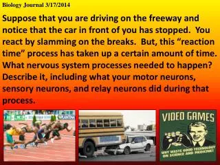
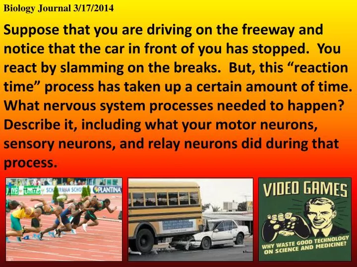
Biology Journal 3/17/2014 Suppose that you are driving on the freeway and notice that the car in front of you has stopped. You react by slamming on the breaks. But, this “reaction time” process has taken up a certain amount of time. What nervous system processes needed to happen? Describe it, including what your motor neurons, sensory neurons, and relay neurons did during that process.
Biology Journal 3/18/2014 The back of your eye is full of specialized neurons called rods and cones. What kind of neuron do you think these cells are? What do you think would be different about their dendrites?
Biology Journal 3/19/2014 What is the most interesting thing you observed from the eye dissection? Dissecting a whale eye.
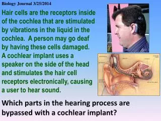
Biology Journal 3/20/2014 What is the name of the nerve that sends signals from the eyes to the brain? The optic nerve What part of the brain processes the signals from the eyes? The primary visual cortex, which is in the back of the brain What part separates the visual signals into the left and right sides? This is called contralateral processing The optic chaism
Label a diagram of the structure of the human eye. The diagram should include: • sclera, cornea, conjunctiva, eyelid, lens, choroid, aqueous humour, pupil, • iris, vitreous humour, retina, fovea, optic nerve, blind spot Eyelid protection, cleaning Choroid layer of light-absorbing pigment Conjunctiva protective outer layer of pupil, secretes mucus Sclera protective outer layer Retina mostly rod cells Fovea area of concentrated cone cells Pupil opening that lets light in Blind Spot no receptor cells Aqueous Humor transparent jelly Lens adjusts to focus light on retina Iris muscles that control size of pupil; gives “eye color” Optic Nerve carries nerve impulses to brain Vitreous humor transparent liquid
What do your pupils do in bright light? What do your pupils do in darkness?
Dilated pupils: when they’re open wide Constricted pupils: when they’re small
Ever have your eyes get sore from staring at a bright computer screen, TV, or phone? That’s because you’re iris muscles are sore from constricting for so long! I’d better check her facebook page again…
Many drugs dilate the pupils because they relax the body’s muscles. This contributes to why vision seems more intense
Nearsighted: The focus of the image falls short of the retina, so its blurry. Farsighted: The focus of the image is beyond the retina, so its blurry.
Rodsrespond to light intensity. They work well in low light. Cones are receptors respond to colors (blue, green, red). They do not work well in low light.
Your peripheral vision has mostly rods. The focus of your retina (called the fovea) contains mostly cones.
How you see: Light stimulates the receptors of the rods and cones. Bipolar cells send the action potential from the rod/cone to the ganglion cells. Ganglion cells collect the action potentials from many rods or cones and send this signal down the retina, toward the optic nerve, where it is sent to the brain.
Action Potential Direction of light Choroid
Cone cell Rod cell Bipolar cell Ganglion cell Direction of nerve signal Direction of light
contralateral processing An image is flipped twice in visual processing: The lens flips it to opposite sides of the retina. The optic chaism flips the image back, where it is carried primary visual cortex (back of the brain) to be interpreted.
Explanation Edge enhancement: Our photoreceptors inhibit neighboring photoreceptors that are the samecolor. Thus, when color borders a different color (there is an “edge”), a photoreceptor won’t be inhibited on the edge, making the edge appear. This helps us to see shapes better. How might edge enhancement help us survive?
Explanation • Retinal Fatigue: Our rods and cones get “fatigued” when being stimulated by the same wavelength of light for a long time, and begin to shut down. • After fatiguing your photoreceptors… • Subtle changes in color get “washed out.” • When you look at a “blank” screen, colors are inverted.
Explanation Retinal Fatigue: Your brain is adapted to reading faces. It does this so well that we often see faces in when there are none. However, his can lead to distressing images when the faces don’t meet our brain’s expectations… Our Devine Savior the Holy Jesus Cheese Sandwich