Download
1 / 26
260 likes | 420 Views
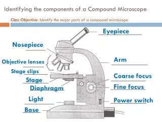
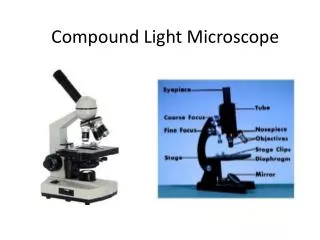
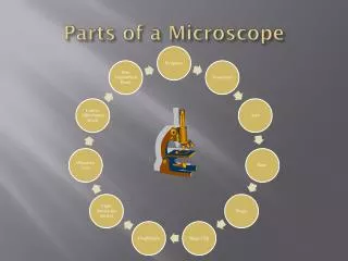
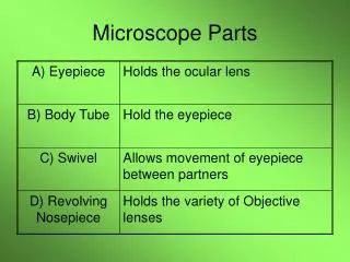
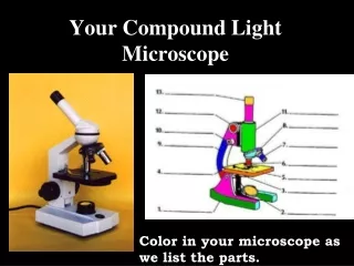
Parts of a Compound Light Microscope. Mirror / Light. the light source for a microscope, typically located in the base of the microscope. Most light microscopes use low voltage, halogen bulbs with continuous variable lighting control located within the base. A. Mirror / Light.

E N D
Mirror / Light • the light source for a microscope, typically located in the base of the microscope. • Most light microscopes use low voltage, halogen bulbs with continuous variable lighting control located within the base.
Coarse adjustment knob • used to focus the microscope • You use it to move the objective lenses toward or away from the specimen.
Fine adjustment knob • used to focus the microscope • This is the knob used to fine tune the focus on the specimen. • It is also used to focus on various parts of the specimen. • Generally one uses the coarse focus first to get close then moves to the fine focus knob for fine tuning.
Eye piece • The eyepiece or Ocular is what you look through at the top of the microscope. • Most eyepieces have a magnifying power of 10x.
Nose piece • The part of the microscope that holds the objective lenses also called a revolving nosepiece or turret. • The objectives are exposed and are mounted so that different objectives can be selected. • Standard objectives include 4x, 10x, 40x and 100x.
Objective lenses • The primary optical lenses on a microscope. • They range from 4x-100x and typically, include, three, four or five on lens on most microscopes.
Stage clips • Holds the slide in place • The viewer is required to move the slide manually to view different sections of the specimen.
Stage • where the specimen to be viewed is placed.
Diaphragm • controls the amount of light reaching the specimen.
Hooke's compound microscope • From "The Microscope and Its Revelations" Carpenter, 1891: image of Hooke's compound microscope 1665
Leeuwenhoek's simple microscope • From "The Microscope and Its Revelations" Carpenter, 1891: images of Leeuwenhoek's simple microscope
Today we will be looking at: • Salt crystals colored thread • Wool fibers newspaper print • Cotton fibers nylon fibers • The letter “e” cork • Dust silk fibers • Volcanic ash corn starch • You will select 5 to practice focusing & drawing (in color) under all three powers
D. Eye piece C. Course adjustment knob E. Nose B. Fine adjustment knob G. Objective lenses H. Stage Clips I. Stage A. Mirror / Light J. Diaphragm