Download
1 / 3

E N D
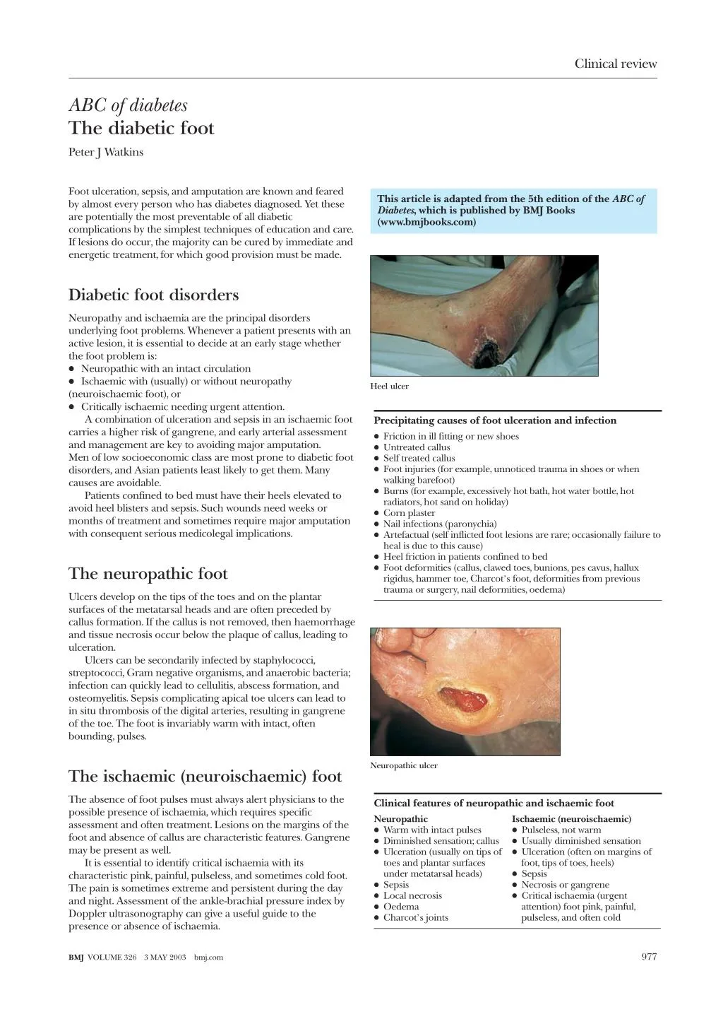
Clinical review ABC of diabetes The diabetic foot Peter J Watkins Foot ulceration, sepsis, and amputation are known and feared by almost every person who has diabetes diagnosed. Yet these are potentially the most preventable of all diabetic complications by the simplest techniques of education and care. If lesions do occur, the majority can be cured by immediate and energetic treatment, for which good provision must be made. This article is adapted from the 5th edition of the ABC of Diabetes, which is published by BMJ Books (www.bmjbooks.com) Diabetic foot disorders Neuropathy and ischaemia are the principal disorders underlying foot problems. Whenever a patient presents with an active lesion, it is essential to decide at an early stage whether the foot problem is: x Neuropathic with an intact circulation x Ischaemic with (usually) or without neuropathy (neuroischaemic foot), or x Critically ischaemic needing urgent attention. A combination of ulceration and sepsis in an ischaemic foot carries a higher risk of gangrene, and early arterial assessment and management are key to avoiding major amputation. Men of low socioeconomic class are most prone to diabetic foot disorders, and Asian patients least likely to get them. Many causes are avoidable. Patients confined to bed must have their heels elevated to avoid heel blisters and sepsis. Such wounds need weeks or months of treatment and sometimes require major amputation with consequent serious medicolegal implications. Heel ulcer Precipitating causes of foot ulceration and infection x Friction in ill fitting or new shoes x Untreated callus x Self treated callus x Foot injuries (for example, unnoticed trauma in shoes or when walking barefoot) x Burns (for example, excessively hot bath, hot water bottle, hot radiators, hot sand on holiday) x Corn plaster x Nail infections (paronychia) x Artefactual (self inflicted foot lesions are rare; occasionally failure to heal is due to this cause) x Heel friction in patients confined to bed x Foot deformities (callus, clawed toes, bunions, pes cavus, hallux rigidus, hammer toe, Charcot’s foot, deformities from previous trauma or surgery, nail deformities, oedema) The neuropathic foot Ulcers develop on the tips of the toes and on the plantar surfaces of the metatarsal heads and are often preceded by callus formation. If the callus is not removed, then haemorrhage and tissue necrosis occur below the plaque of callus, leading to ulceration. Ulcers can be secondarily infected by staphylococci, streptococci, Gram negative organisms, and anaerobic bacteria; infection can quickly lead to cellulitis, abscess formation, and osteomyelitis. Sepsis complicating apical toe ulcers can lead to in situ thrombosis of the digital arteries, resulting in gangrene of the toe. The foot is invariably warm with intact, often bounding, pulses. Neuropathic ulcer The ischaemic (neuroischaemic) foot The absence of foot pulses must always alert physicians to the possible presence of ischaemia, which requires specific assessment and often treatment. Lesions on the margins of the foot and absence of callus are characteristic features. Gangrene may be present as well. It is essential to identify critical ischaemia with its characteristic pink, painful, pulseless, and sometimes cold foot. The pain is sometimes extreme and persistent during the day and night. Assessment of the ankle-brachial pressure index by Doppler ultrasonography can give a useful guide to the presence or absence of ischaemia. Clinical features of neuropathic and ischaemic foot Neuropathic x Warm with intact pulses x Diminished sensation; callus x Ulceration (usually on tips of toes and plantar surfaces under metatarsal heads) x Sepsis x Local necrosis x Oedema x Charcot’s joints Ischaemic (neuroischaemic) x Pulseless, not warm x Usually diminished sensation x Ulceration (often on margins of foot, tips of toes, heels) x Sepsis x Necrosis or gangrene x Critical ischaemia (urgent attention) foot pink, painful, pulseless, and often cold 977 BMJ VOLUME 326 3 MAY 2003 bmj.com
Clinical review Management Specialist care of the ulcerated foot Expect the team to ensure as a minimum: x Local wound management, appropriate dressings, and debridement as indicated x Antibiotic treatment as appropriate x Investigation and management of vascular insufficiency x Specialist footwear to distribute foot pressures appropriately x Good blood glucose control Infected diabetic foot lesions should be treated only by those with sufficient experience and facilities. General practitioners rarely have such experience and should normally refer patients for urgent specialist care from the foot care team. Treatment of diabetic foot ulcers Management of ulcers falls into three parts: removal of callus, eradication of infection, and reduction of weight bearing forces, often requiring bed rest with the foot raised. Excess keratin should be pared away with a scalpel blade to expose the floor of the ulcer and allow efficient drainage of the lesion. A radiograph should be taken to look for osteomyelitis whenever a deep penetrating ulcer is present or when lesions fail to heal or continue to recur. A bacterial swab should be taken from the floor of the ulcer after the callus has been removed; culture of excised tissue may yield even more reliable information. Patients with superficial ulcers can be treated as outpatients and prescribed appropriate oral antibiotics until the ulcer has healed. The most likely organisms to infect a superficial ulcer are staphylococci, streptococci, and sometimes anaerobes. Thus, treatment is started with amoxicillin, flucloxacillin, and metronidazole and adjusted when the results of bacteriological culture are available. Choice and duration of antibiotic treatment require considerable expertise and laboratory guidance. The patient should be instructed to dress the ulcer daily. A simple non-adherent dressing should be applied after cleaning the ulcer with physiological saline. Deep indolent ulcers also require local wound care and antibiotics. Application of a total contact plaster cast, lightweight scotch cast boot, or air cast boots may help healing. These conform to the contours of the foot, thereby reducing shear forces on the plantar surface. Great care must be taken, especially with the fitting of plasters, to prevent chafing and subsequent ulcer formation elsewhere on the foot or ankle. Any foot lesion that has not healed in one month requires further investigation and a different approach. Radiograph showing osteomyelitis Ischaemic ulcer Danger signs: urgent treatment needed x Redness and swelling of a foot that even when neuropathic causes some discomfort and pain; this often indicates a developing abscess, and urgent surgery may be needed to save the leg x Cellulitis, discolouration, and crepitus (gas in soft tissues) x Pink, painful, pulseless foot even without gangrene indicates critical ischaemia that needs urgent arterial investigation followed by surgical intervention whenever possible Urgent treatment Patients with the danger signs listed in the box need to be admitted to hospital immediately for urgent treatment and investigation. They should have bed rest and be started on intravenous antibiotics straight away. An intravenous insulin pump may be needed to control blood glucose concentration. Antibiotics—In the first 24 hours before bacteriological cultures are available, a wide spectrum of antibiotic cover is needed. Quadruple therapy may be necessary, consisting of amoxicillin, flucloxacillin with metronidazole (to treat anaerobes), and either ceftazidime 1 g three times daily or gentamicin to treat Gram negative organisms. This treatment can be adapted when the results of bacteriological culture are available. The emergence of multiple resistant Staphylococcus aureus (MRSA) is presenting a serious problem, firstly, because it can be responsible for the ravages of sepsis and, secondly, because these patients need isolation while in hospital. Available treatments include intravenous vancomycin and intramuscular teicoplanin. Surgical debridement is needed to drain pus and abscess cavities and to remove all necrotic and infected tissue including devitalised and infected bone resulting from osteomyelitis. Deep tissue swabs should be sent to the laboratory. If necrosis has developed in the digit, a ray amputation to remove the toe and part of its associated metatarsal is necessary and is usually successful in the neuropathic foot with intact circulation. Skin grafting is occasionally needed and accelerates wound healing. Critical ischaemia with ischaemic gangrene of great toe 978 BMJ VOLUME 326 3 MAY 2003 bmj.com
Clinical review The ischaemic foot Sepsis in the presence of ischaemia is a dangerous combination and should be treated urgently as described above. When ischaemia is suspected, or an ulcer does not respond to medical treatment, vascular investigation is required. Doppler studies to measure the ankle-brachial pressure index (the ratio of systolic blood pressure at the ankle and brachial artery) give valuable information about the state of the vessels. Arterial imaging by duplex scanning, magnetic resonance angiography, or conventional arteriography is done with a view to angioplasty, arterial reconstruction, or both. Infrapopliteal angioplasty or distal bypass to the tibial or peroneal vessels are now well established and are important for limb salvage. The intravenous dye used during angiography can precipitate acute oliguric renal failure in patients with early renal impairment. Preventative measures must therefore be taken. Amputation of the toe is usually unsuccessful unless the foot can be revascularised. If this is not possible, a dry necrotic toe should be allowed to autoamputate. After attempts to control infection, below knee amputation is indicated in patients with rampant progressive infection or extensive tissue destruction. Rest pain in the severely ischaemic limb may be relieved by revascularisation, but if that fails, pain relief with opiates may be necessary. Paravertebral lumbar block has been disappointing in promoting healing, but occasionally rest pain is ameliorated. If all these measures fail and pain remains intractable, then below knee amputation may be needed. Interpreting the ankle-brachial pressure index Pressure index >1.2 >1 <0.9 <0.6 State of vessels Rigid or calcified vessels or both Normal (or calcified) Ischaemia Severe ischaemia Note: Vascular calcification is common so spuriously high readings can be obtained. This must be taken into account when the pressure index reading is evaluated. Renal protection during arteriography x Avoid dehydration x Give intravenous fluids, starting four hours before the procedure x Use intravenous insulin sliding scale x Monitor urine output x Check creatinine concentrations before and on the day after the procedure Note: Metformin should be stopped 48 hours before angiography and resumed 48 hours after the procedure has been completed. The neuropathic joint (Charcot’s joint) Loss of pain sensation together with possible rarefaction of the bones of the neuropathic foot can have serious consequences. Abnormal mechanical stresses that are usually prevented by pain may occur, and the susceptible bones are then damaged by relatively minor trauma. Patients present with a hot swollen foot, sometimes aching, and the appearances are often mistaken for infection. Injury may have occurred days or weeks earlier, or may not even have been noticed. Sometimes Charcot changes develop after minor amputations that alter the normal weight bearing stresses. The destructive process does not continue indefinitely but stops after weeks or months. Bony changes are most common at the tarsal-metatarsal region of the foot but also occur at the ankle and metatarso-phalangeal region. Early diagnosis is essential. Unilateral warmth and swelling in a neuropathic foot is suggestive of a developing Charcot’s joint. Bone scans are more sensitive indicators of new bone formation than radiography and should be used to confirm the diagnosis. Gallium white cell scanning and magnetic resonance imaging should be done to exclude infection as the cause. Management initially comprises rest, ideally bed rest or use of non-weight bearing crutches, until the oedema and local warmth have resolved. Alternatively, the foot can be immobilised in a well moulded total contact plaster that is initially non-weight bearing. Immobilisation is continued until bony repair is complete (usually 2-3 months). The use of bisphosphonates to prevent bone damage in Charcot’s foot is being investigated and seems promising. Long term, special shoes and insoles should be fitted to accommodate deformity and prevent ulceration. Neuropathic Charcot’s joint Isotope bone scans of a normal foot (left) and a neuropathic foot (right) showing high blood flow Long term care after wound healing is complete Appropriate footwear Ongoing podiatry to remove excess callus and provide nail care Regular assessment: x Identify critically ischaemic foot x Look for active lesions (for example, hidden between the toes) and treat immediately x Detect and manage deformities, callus, skin cracks, and discoloration x Simple sensory test such as monofilament sensory test under the great toe (inability to detect ≥10 g indicates risk of foot ulceration) x Examine pulses (dorsalis pedis and posterior tibial) x Assess ankle reflex x Assess other sensory modalities—for example, pinprick and vibration perception at the medial malleolus or tip of the great toe Neuropathic oedema Neuropathic oedema consists of swelling of the feet and lower legs associated with severe peripheral neuropathy; it is uncommon. The pathogenesis may be related to vasomotor changes and arteriovenous shunting. Ephedrine 30 mg three times daily reduces peripheral oedema by reducing blood flow and increasing sodium excretion. Peter J Watkins is honorary consultant physician, King’s Diabetes Centre, King’s College Hospital, London (peter.watkins1@virgin.net). BMJ 2003;326:977–9 979 BMJ VOLUME 326 3 MAY 2003 bmj.com