Download
1 / 1
10 likes | 142 Views
Affinity Protein Separation by Pulse Electrophoresis in Functionalized Anodic Aluminum Oxide (AAO) Membranes. Zhiqiang Chen, Tao Chen , Xinghua Sun , Bruce. J. Hinds*. Department of Chemical and Materials Engineering, University of Kentucky, Lexington, KY 40506. Introduction.

E N D
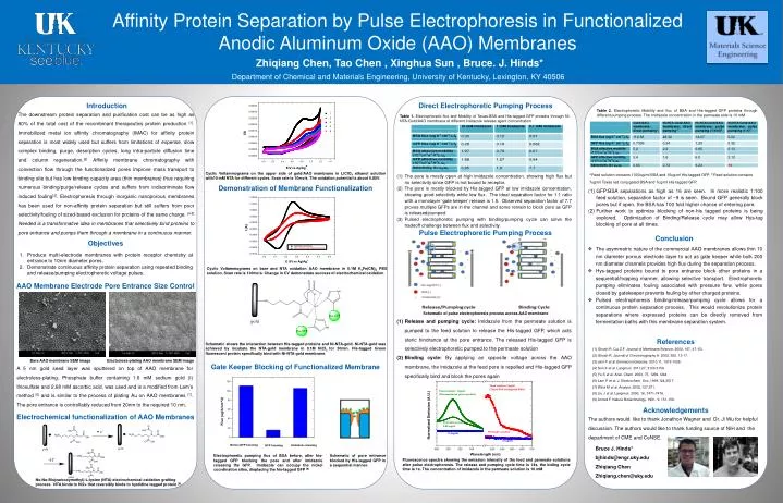
Affinity Protein Separation by Pulse Electrophoresis in Functionalized Anodic Aluminum Oxide (AAO) Membranes Zhiqiang Chen, Tao Chen , Xinghua Sun , Bruce. J. Hinds* Department of Chemical and Materials Engineering, University of Kentucky, Lexington, KY 40506 Introduction Direct Electrophoretic Pumping Process Table 2. Electrophoretic Mobility and flux of BSA and His-tagged GFP proteins through different pumping process. The imidazole concentration in the permeate side is 10 mM The downstream protein separation and purification cost can be as high as 80% of the total cost of the recombinant therapeutics protein production [1].Immobilized metal ion affinity chromatography (IMAC) for affinity protein separation is most widely used but suffers from limitations of expense, slow complex binding, purge, desorption cycles, long intra-particle diffusion time and column regeneration.[2] Affinity membrane chromatography with convection flow through the functionalized pores improve mass transport to binding site but has low binding capacity area (thin membranes) thus requiring numerous binding/purge/release cycles and suffers from indiscriminate flow induced fouling[3]. Electrophoresis through inorganic nanoporous membranes has been used for non-affinity protein separation but still suffers from poor selectivity/fouling of sized-based exclusion for proteins of the same charge. [4-5] Needed is a transformative idea in membranes that selectively bind proteins to pore entrance and pumps them through a membrane in a continuous manner. Table 1. Electrophoretic flux and Mobility of Texas-BSA and His-tagged GFP proteins through Ni-NTA-Gold/AAO membrane at different imidazole release agent concentration Cyclic Voltammograms on the upper side of gold/AAO membrane in LiClO4 ethanol solution with10 mM NTA for different cycles. Scan rate is 10mv/s. The oxidation potential is about 0.85V. a Feed solution contains 1000ug/ml BSA and 10ug/ml His-tagged GFP. b Feed solution contains 7ug/ml Texas red conjugated BSA and 7ug/ml His-tagged GFP. The pore is mostly open at high imidazole concentration, showing high flux but no selectivity since GFP is not bound to receptor. The pore is mostly blocked by His-tagged GFP at low imidazole concentration, showing good selectivity while low flux. The ideal separation factor for 1:1 ratio with a monolayer ‘gate keeper’ release is 1.5. Observed separation factor of 7.7 proves multiple GFPs are in the channel and some remain to block pore as GFP is released/pumped. Pulsed electrophoretic pumping with binding/pumping cycle can solve the tradeoff challenge between flux and selectivity. Demonstration of Membrane Functionalization GFP:BSA separations as high as 16 are seen. In more realistic 1:100 feed solution, separation factor of ~9 is seen. Bound GFP generally block pores but if open, the BSA has 100 fold higher chance of entering pore. Further work to optimize blocking of non-his tagged proteins is being explored. Optimization of Binding/Release cycle may allow Hys-tag blocking of pore at all times. Pulse Electrophoretic Pumping Process Conclusion Objectives • The asymmetric nature of the commercial AAO membranes allows thin 10 nm diameter porous electrode layer to act as gate keeper while bulk 200 nm diameter channels provides high flux during the separation process. • Hys-tagged proteins bound to pore entrance block other proteins in a sequential/hopping manner, allowing selective transport. Electrophoretic pumping eliminates fouling associated with pressure flow, while pores closed by gatekeeper prevents fouling by other charged proteins. • Pulsed electrophoresis binding/release/pumping cycle allows for a continuous protein separation process. This would revolutionize protein separations where expressed proteins can be directly removed from fermentation baths with this membrane separation system. Produce multi-electrode membranes with protein receptor chemistry at entrance to 10nm diameter pores. Demonstrate continuous affinity protein separation using repeated binding and release/pumping electrophoretic voltage pulses. Cyclic Voltammograms on bare and NTA oxidation AAO membrane in 0.1M K3Fe(CN)6 PBS solution. Scan rate is 100mv/s. Change in CV demonstrate success of electrochemical oxidation AAO Membrane Electrode Pore Entrance Size Control Schematic of pulse electrophoresis process across AAO membrane • Release and pumping cycle: Imidazole from the permeate solution is pumped to the feed solution to release the His-tagged GFP, which acts steric hindrance at the pore entrance. The released His-tagged GFP is selectively electrophoretic pumped to the permeate solution • Binding cycle: By applying an opposite voltage across the AAO membrane, the imidazole at the feed pore is repelled and His-tagged GFP specifically bind and block the pores again References Schematic shows the interaction between His-tagged proteins and Ni-NTA-gold. Ni-NTA-gold was achieved by incubate the NTA-gold membrane in 0.1M NiCI2 for 30min. His-tagged Green fluorescent protein specifically bind with Ni-NTA-gold membrane. Ghosh R, Cui Z.F. Journal of Membrane Science. 2000, 167, 47–53. Ghosh R. Journal of Chromotography A. 2002, 952, 13-17. Jain P et al. Biomacromolecules. 2010,11, 1019-1026. Sun X et al. Langmuir. 2011,27, 3150-3156. Yu S et al. Anal. Chem. 2003, 75, 1239-1244 Lam P et al. J. Electrochem. Soc.,1999,124,2517. Wirtz M et al. Analyst. 2002, 127,871. Liu J et al. Langmuir. 2000, 16, 7471-7476. Arnold F. Nature Biotechnology. 1991, 9, 151-156. Bare AAO membrane SEM image Electroless-plating AAO membrane SEM image Gate Keeper Blocking of Functionalized Membrane A 5 nm gold seed layer was sputtered on top of AAO membrane for electroless-plating. Phosphate buffer containing 1.6 mM sodium gold (I) thiosulfate and 2.68 mM ascorbic acid, was used and is a modified from Lam’s method [6] and is similar to the process of plating Au on AAO membranes [7]. The pore entrance is controllably reduced from 20nm to the required 10 nm. Acknowledgements Electrochemical functionalization of AAO Membranes The authors would like to thank Jonathon Wagner and Dr. Ji Wu for helpful discussion. The authors would like to thank funding source of NIH and the department of CME and CeNSE. Bruce J. Hinds* bjhinds@engr.uky.edu Zhiqiang Chen Zhiqiang.chen@uky.edu Electrophoretic pumping flux of BSA before, after his-tagged GFP blocking the pore and after imidazole releasing the GFP. Imidazole can occupy the nickel coordination sites, displacing the his-tagged GFP [9] Schematic of pore entrance blocked by His-tagged GFP in a sequential manner. Fluorescence spectra showing the emission intensity of the feed and permeate solutions after pulse electrophoresis. The release and pumping cycle time is 14s, the biding cycle time is 1s. The concentration of imidazole in the permeate solution is 10 mM Nα,Nα-Bis(carboxymethyl)-L-lysine (NTA) electrochemical oxidation grafting process. NTA binds to Ni2+ that reversibly binds to hystidine tagged protein [8]