Download
1 / 1
10 likes | 102 Views
Proteomic Analysis of Skeletal Framework Proteins from the Stony Coral, Stylophora pistillata Jeana L. Drake, Tali Mass, Liti Haramaty , Ehud Zelzion , Debashish Bhattacharya, Paul Falkowski Institute of Marine and Coastal Sciences, Rutgers University, New Brunswick, NJ.

E N D
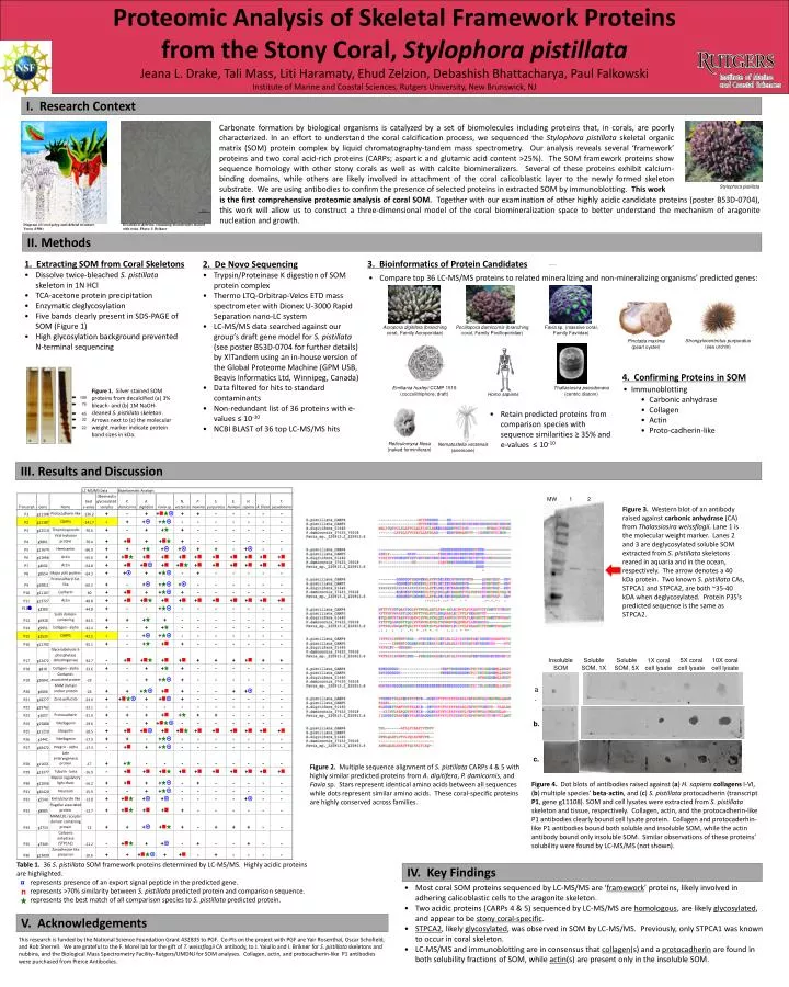
Proteomic Analysis of Skeletal Framework Proteins from the Stony Coral, Stylophorapistillata Jeana L. Drake, Tali Mass, LitiHaramaty, Ehud Zelzion, Debashish Bhattacharya, Paul Falkowski Institute of Marine and Coastal Sciences, Rutgers University, New Brunswick, NJ I. Research Context Carbonate formation by biological organisms is catalyzed by a set of biomolecules including proteins that, in corals, are poorly characterized. In an effort to understand the coral calcification process, we sequenced the Stylophorapistillata skeletal organic matrix (SOM) protein complex by liquid chromatography-tandem mass spectrometry. Our analysis reveals several ‘framework’ proteins and two coral acid-rich proteins (CARPs; aspartic and glutamic acid content >25%). The SOM framework proteins show sequence homology with other stony corals as well as with calcite biomineralizers. Several of these proteins exhibit calcium-binding domains, while others are likely involved in attachment of the coral calicoblastic layer to the newly formed skeleton substrate. We are using antibodies to confirm the presence of selected proteins in extracted SOM by immunoblotting. This work Stylophorapistillata is the first comprehensive proteomic analysis of coral SOM. Together with our examination of other highly acidic candidate proteins (poster B53D-0704), this work will allow us to construct a three-dimensional model of the coral biomineralization space to better understand the mechanism of aragonite nucleation and growth. Diagram of coral polyp and skeletal structure. Veron(1986) Decalcified skeleton; remaining biomolecules stained with eosin. Photo: I. Brikner II. Methods 3. Bioinformatics of Protein Candidates • 1. Extracting SOM from Coral Skeletons • Dissolve twice-bleached S. pistillataskeleton in 1N HCl • TCA-acetone protein precipitation • Enzymatic deglycosylation • Five bands clearly present in SDS-PAGE of SOM (Figure 1) • High glycosylation background prevented N-terminal sequencing • 2. De Novo Sequencing • Trypsin/Proteinase K digestion of SOM protein complex • Thermo LTQ-Orbitrap-Velos ETD mass spectrometer with Dionex U-3000 Rapid Separation nano-LC system • LC-MS/MS data searched against our group’s draft gene model for S. pistillata(see poster B53D-0704 for further details) by X!Tandemusing an in-house version of the Global Proteome Machine (GPM USB, Beavis Informatics Ltd, Winnipeg, Canada) • Data filtered for hits to standard contaminants • Non-redundant list of 36 proteins with e-values ≤ 10-10 • NCBI BLAST of 36 top LC-MS/MS hits • Compare top 36 LC-MS/MS proteins to related mineralizing and non-mineralizing organisms’ predicted genes: • Acroporadigitifera(branching coral, Family Acroporidae) • Favia sp. (massive coral, Family Faviidae) • Pocilloporadamicornis(branching coral, Family Pocilloporidae) • Strongylocentrotuspurpuratus (sea urchin) • Pinctada maxima (pearl oyster) 4. Confirming Proteins in SOM • Immunoblotting • Carbonic anhydrase • Collagen • Actin • Proto-cadherin-like • EmilianiahuxleyiCCMP 1516 (coccolithiphore; draft) • Thallasiosirapseudonana(centric diatom) Figure 1. Silver stained SOM proteins from decalcified (a) 3% bleach- and (b) 1M NaOH-cleaned S. pistillataskeleton. Arrows next to (c) the molecular weight marker indicate protein band sizes in kDa. • Homo sapiens • Retain predicted proteins from comparison species with sequence similarities ≥ 35% and e-values ≤ 10-10 • Reticulomyxafilosa(naked forminiferan) • Nematostellavectensis(anemone) III. Results and Discussion 2 MW 1 Figure 3. Western blot of an antibody raised against carbonic anhydrase (CA) from Thalassiosiraweissflogii. Lane 1 is the molecular weight marker. Lanes 2 and 3 are deglycosylated soluble SOM extracted from S. pistillataskeletons reared in aquaria and in the ocean, respectively. The arrow denotes a 40 kDa protein. Two known S. pistillataCAs, STPCA1 and STPCA2, are both ~35-40 kDA when deglycosylated. Protein P35’s predicted sequence is the same as STPCA2. Insoluble SOM Soluble SOM, 1X Soluble SOM, 5X 5X coral cell lysate 10X coral cell lysate 1X coral cell lysate a. b. c. Figure 2. Multiple sequence alignment of S. pistillataCARPs 4 & 5 with highly similar predicted proteins from A. digitifera, P. damicornis, and Favia sp. Stars represent identical amino acids between all sequences while dots represent similar amino acids. These coral-specific proteins are highly conserved across families. Figure 4. Dot blots of antibodies raised against (a) H. sapiens collagens I-VI, (b) multiple species’ beta-actin, and (c) S. pistillataprotocadherin (transcript P1, gene g11108). SOM and cell lysates were extracted from S. pistillata skeleton and tissue, respectively. Collagen, actin, and the protocadherin-like P1 antibodies clearly bound cell lysate protein. Collagen and protocaderhin-like P1 antibodies bound both soluble and insoluble SOM, while the actin antibody bound only insoluble SOM. Similar observations of these proteins’ solubility were found by LC-MS/MS (not shown). Table 1. 36 S. pistillataSOM framework proteins determined by LC-MS/MS. Highly acidic proteins are highlighted. This represents presence of an export signal peptide in the predicted gene. This represents >70% similarity between S. pistillatapredicted protein and comparison sequence. This represents the best match of all comparison species to S. pistillatapredicted protein. IV. Key Findings • Most coral SOM proteins sequenced by LC-MS/MS are ‘framework’ proteins, likely involved in adhering calicoblastic cells to the aragonite skeleton. • Two acidic proteins (CARPs 4 & 5) sequenced by LC-MS/MS are homologous, are likely glycosylated, and appear to be stony coral-specific. • STPCA2, likely glycosylated, was observed in SOM by LC-MS/MS. Previously, only STPCA1 was known to occur in coral skeleton. • LC-MS/MS and immunoblotting are in consensus that collagen(s) and a protocadherin are found in both solubility fractions of SOM, while actin(s) are present only in the insoluble SOM. V. Acknowledgements This research is funded by the National Science Foundation Grant 432835 to PGF. Co-PIs on the project with PGF are Yair Rosenthal, Oscar Schofield, and Rob Sherrell. We are grateful to the F. Morel lab for the gift of T. weissflogiiCA antibody, to J. Yaiullo and I. Brikner for S. pistillata skeletons and nubbins, and the Biological Mass Spectrometry Facility-Rutgers/UMDNJ for SOM analyses. Collagen, actin, and protocadherin-like P1 antibodies were purchased from Pierce Antibodies.