Download
1 / 30
300 likes | 479 Views
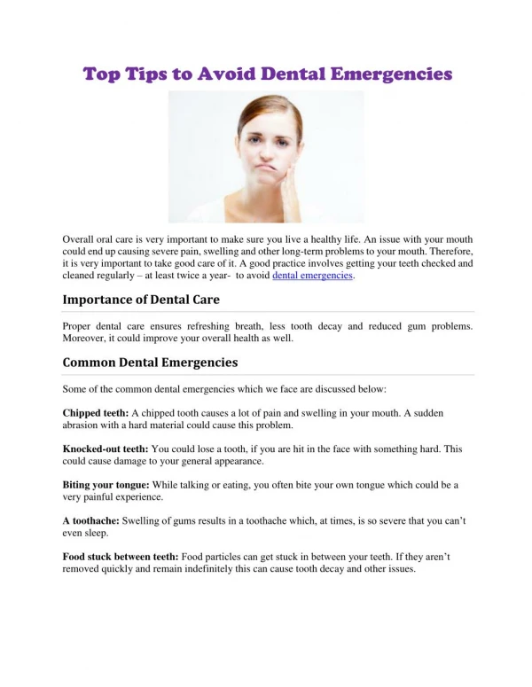
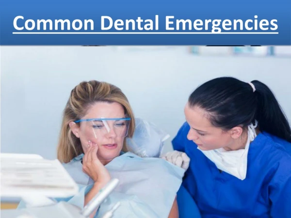
Dental and Ophthalmologic Emergencies. Morehouse Pediatrics EM Lecture Series November 23,2009 Taryn R Taylor, MD. Dental Emergencies. Epidemiology Tooth Eruption & Shedding Schedule Clinical Evaluation Dental Concussion & Subluxation Avulsion Injuries Tooth Displacement Tooth Fractures

E N D
Dental and Ophthalmologic Emergencies Morehouse Pediatrics EM Lecture Series November 23,2009 Taryn R Taylor, MD
Dental Emergencies • Epidemiology • Tooth Eruption & Shedding Schedule • Clinical Evaluation • Dental Concussion & Subluxation • Avulsion Injuries • Tooth Displacement • Tooth Fractures • Dental Abscesses
Dental Emergencies • Epidemiology • 30% of children experience dental injuries • Peak period of trauma to primary teeth is 18 to 40 months of age • Trauma to permanent teeth • School aged boys suffer trauma twice as frequently as girls • Upper (maxillary) central incisors are most frequently injured
Dental Emergencies • Tooth Eruption & Shedding Schedule
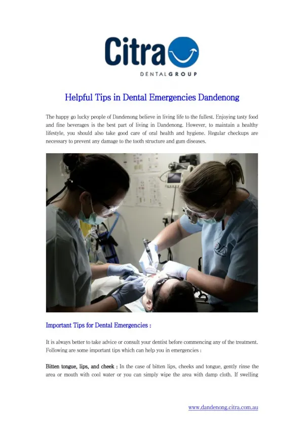
Dental Emergencies • Clinical Evaluation • Medical History • Assess need for SBE prophylaxis • Determine if child has a bleeding disorder or is immunocompromised • Record current medications and medication allergies • Obtain history of previous surgeries • Determine if tetanus immunization is up to date • Determine if child lost consciousness due to injury • Dental History • How the injury occurred: provides info regarding severity • When injury occurred: prognosis for injured tooth worsens with every minute of delay in treatment • Where injury occurred: helps determine whether tetanus prophylaxis is warranted
Dental Emergencies • Clinical Evaluation • Physical Examination • General assessment includes review of vital signs, evaluation of potential head and cervical spine injury as well as ocular damage • Extra oral evaluation • Palpate mandibular condyles, maxilla, zygoma & TMJ • Anterior open bite, malocclusion or limited mandibular opening suggests condylar fractures or dislocation • Note extra oral lacerations, bruises or swelling • Lacerations must be inspected for foreign bodies i.e. gravel or tooth fragments & be debrided if foreign body present
Dental Emergencies • Physical Exam cont. • Intra oral evaluation • Remove all clots and debris • Palpate alveolus to detect fractures • Have patient clench teeth to detect dental occlusion • Examine each tooth for damage or mobility • Examine labial mucosa, maxillary frenulum, gingival tissues and tongue for bruising or lacerations • Lacerations must be cleaned & explored for presence of foreign body • Frenulum will heal without long term consequences • Most tongue lacerations will heal on their own, unless tissue edges are not self-approximating
Dental Emergencies • Dental Concussion & Subluxation • Concussion: Mild injury to periodontal ligament without tooth mobility or displacement • Subluxation: Significant injury to periodontal ligament resulting in some tooth mobility • These injuries may result in tooth discoloration • Initial management • Tylenol as needed for pain • Ice as needed for swelling • Soft diet • Follow up with dentist • Dental office management • Radiographs of primary tooth to evaluate for root fracture • Splinting of permanent tooth if extremely mobile
Dental Emergencies • Avulsion Injuries • Occurs when a tooth is completely displaced from the dental socket • Radiographs may be necessary if tooth cannot be found • Primary Teeth • Not reimplanted, as the risk of injury to developing permanent tooth bud is high • Permanent Teeth • Best way to preserve an avulsed tooth is to replace it in its socket as quickly as possible • Periodontal ligament is protective layer surrounding the root, which suffers irreversible damage if allowed to dry • Do not touch root of tooth, handle by crown only • Rinse only if there is dirt covering it, don’t scrub or scrape tooth • Gently dislodge any clots, & reintroduce tooth into the dental socket slowly
Dental Emergencies • Avulsion Injuries cont. • Post Reimplantation care • Dental consult immediately for splinting & tooth stabilization • 10 day course of prophylactic penicillin • Tetanus vaccination if wound is dirty or vaccination requires updating • Chlorhexidine gluconate rinses, oral hygiene & soft diet instructions • Analgesics for pain control • Dental follow up within one week
Dental Emergencies • Tooth Displacement • Luxation: Displacement of tooth in any direction, while remaining in the socket • Lateral luxation is usually associated with fracture of alveolar bone • Primary teeth: analgesia, proper dental hygiene, prompt dentist follow • Permanent teeth: gently reposition tooth, additional care similar to primary teeth
Dental Emergencies • Tooth Displacement • Extrusion: tooth is only partially removed from socket • Care similar to other luxation injuries • Intrusion: tooth is impacted into alveolar bone with associated fracture • Intrusions of up to 3 mm have excellent prognosis • Care similar to other luxation injuries
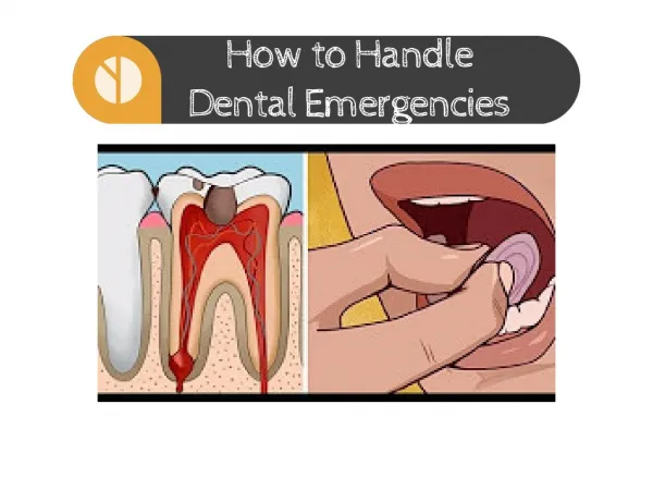
Dental Emergencies • Tooth Fractures • Crown fractures are described by Ellis Classification • Ellis class I: involves enamel only, rarely painful, cosmetic implications only • Ellis class II: involves enamel as well as dentin • Sensitivity to cold air & fluids • Emergency treatment aimed at protecting the pulp by applying calcium hydroxide product • Dentist follow up in 48 hours • Ellis class III: dental pulp involved, often appears red • Exposure of nerve endings causes extreme pain • Exposure of pulp will lead to pulpal necrosis from bacterial infection if left untreated • Emergency treatment aimed at protecting the pulp by applying calcium hydroxide product • Dentist follow up within 24 hours
Dental Emergencies • Dental Abscess • Results when inflammation of the pulp is left untreated • Pain, tenderness, red, swollen gingiva with areas of fluctuance • Complications include localized cellulitis, fistula formation • Emergency management includes pain control and oral penicillin • Prompt dental follow-up • Emergent ENT consultation for patients requiring incision and drainage due to severe pain or with extension of infection into deeper tissues
Ophthalmologic Emergencies • Landmarks of the eye • Different types of eye injuries • Emergency care for eye injuries • Orbital Cellulitis
Ophthalmologic Emergencies • The globe of the eye, or eyeball is a sphere approximately 1” in diameter • Five most important landmarks of the eye: • Sclera- the “white” of the eye • Cornea- clear, front portion of the eye that covers the pupil • Pupil- opening in which light enters • Iris-colored portion of the eye • Retina- back of the eye
Ophthalmologic Emergencies • Ocular trauma is the leading cause of noncongenital unilateral blindness in children younger than 20 • Most eye trauma occurs during sports activities • Clinical Assessment • Mechanism: blunt or sharp object, foreign body present • Symptoms: pain, photophobia, eye movements, visual acuity • Exam: Pupil size, shape, reaction to light: orbital rims, floor, extra ocular motion
Ophthalmologic Emergencies • Eye injuries are usually not life-threatening • Time is of the essence in your treatment • Six different types of eye injuries: • Foreign object in the eye • Corneal Abrasions • Lid injury • Injury to the globe • Injury to the orbits • Chemical burn to the eye
Ophthalmologic Emergencies • Extra ocular Foreign Objects • Dust, dirt, sand or fine pieces of metal can be blown into the eye & lodged on conjunctiva or cornea • Signs & Symptoms • Pain, foreign body sensation • Excessive tearing • Reddening of conjunctiva • Decreased visual acuity
Ophthalmologic Emergencies • Extra ocular Foreign Body • Flush eye for at least 20 minutes • If object cannot be flushed, attempt to remove • Evaluate for possible corneal abrasion • To remove object: • Pull down lower lid while patient looks up, or evert upper lid while patient looks down • Remove object with sterile gauze
Ophthalmologic Emergencies • Corneal Abrasions • Most common eye injury in all ages • Scraping away of the corneal surface, caused by: • Injury • Blowing dust, sand, debris • Extended contact lens wear • Ocular foreign bodies, embedded under an eyelid • Signs and symptoms • Red, irritated eye • Foreign body sensation • Increased tearing • Photophobia • Fluorescein uptake under Woods lamp • Treatment • Polytrim antibiotic ointment or gtts
Ophthalmologic Emergencies • Eyelid Lacerations • Control bleeding with LIGHT pressure • Ocular injury should always be suspected • Lids should be everted and conjunctival surface examined • Orbital CT if suspected ocular penetration • Laceration repair with 6-0 nonabsorbable suture • Optho referral for repair: • Lacerations involving nasolacrimal duct • Full thickness lacerations • Eyelid margin lacerations • Lacerations from animal or human bites require tetanus prophylaxis
Ophthalmologic Emergencies • Injuries to Globe • Subconjunctival Hemorrhage • Blood between conjunctiva & sclera, stops at cornea • Not an emergency • Heals like any other bruise • Hyphema • Accumulation of blood in the anterior chamber • Complications include inflammation and increased IOP • Patients with sickle cell disease or trait & thalassemia are at risk for central retinal artery and optic nerve damage • Patients are at risk for rebleeding 3-5 days after initial injury • Initial treatment: bed rest, elevation of head of bed 30 degrees, optho referral • Hyphemas > 50% should be admitted • Avoid aspirin & NSAIDS
Ophthalmologic Emergencies • Injuries to Globe • Globe Rupture • Can occur after significant laceration of cornea or sclera due to sharp objects, or blunt trauma • Visual loss, bloody chemosis, soft globe • Protective shield should be placed over the eye AVOIDING direct pressure on globe • Broad-spectrum IV antibiotics against skin flora & tetanus prophylaxis should be administered • Analgesics, sedatives and antiemetics to decrease IOP from vomiting • Immediate ophthalmologic consultation required
Ophthalmologic Emergencies • Orbital Fractures • “Blowout” Fracture • Following blunt trauma, eye is pushed through floor of orbit, causing fracture of orbital wall • Trapping of intraocular muscle prevents movement of eye away from fracture site • Facial asymmetry, sunken eye, paralysis of upwards gaze, double vision • Orbital roof fractures • Occur mostly in children under 5 years • Possibility of communication between orbit & intracranial cavity • Pulsating proptosis • CT scan with immediate ophthalmologic consultation
Ophthalmologic Emergencies • Chemical Burns • Represent a DIRE emergency • Permanent damage can occur within seconds • Burning and tissue damage will continue to occur as long as substance is left in eye • Signs and Symptoms • Irritated, swollen eyelids • Redness of the eye • Blurred/diminished vision • Irritated, burned skin around the eyes
Ophthalmologic Emergencies • Chemical Burns • Emergency Care • Immediately begin irrigation with NS or LR • Continuously irrigate for a minimum of 20 minutes • Remove contact lenses-may trap chemicals • Wash your hands afterward to prevent contamination to yourself • Contact Poison Control Center for further information • Referral to ophthalmologist
Ophthalmologic Emergencies • Orbital Cellulitis • An infection of the orbit itself, which occurs • As a complication of sinusitis with extension of the infection to the orbit • Secondary to penetrating trauma • As an extension of a nearby facial infection • Signs & Symptoms • Erythema, edema, induration and tenderness to peri orbital tissues • Decreased eye movement • Proptosis, chemosis, decreased visual acuity and papilledema
Ophthalmologic Emergencies • Orbital cellulitis • CT scan of orbit • Prompt treatment with IV antibiotics • Inpatient admission for frequent monitoring • Ophthalmologic consultation