Download
1 / 19
200 likes | 321 Views
cell division: mitosis. biology 1. Why cells divide and what they have to do to accomplish this Division in prokaryotes Division in eukaryotes Chromosone structure Phases of the cell cycle Cell growth factors Cancerous growths. Why divide?. Ensures persistence of genome

E N D
cell division: mitosis biology 1
Why cells divide and what they have to do to accomplish this • Division in prokaryotes • Division in eukaryotes • Chromosone structure • Phases of the cell cycle • Cell growth factors • Cancerous growths
Why divide? • Ensures persistence of genome • Precisely replicates DNA • Equally distributes DNA to opposite end of cell • Seperates into two identical daughter cells • Strategy to counter loss of SA:Vol ratio as cell grows larger • Permits growth and development of a multicellular organism • Allows replacement of damaged or dead cells Genome = total endowment of DNA unique to each species
Meiosis in the gonads halves the chromosone number Individual inherits 46 chromosones, 23 from each parent Sperm cell (23 chromosones) Ovum (23 chromosones) Mitosis produces genetically identical daughter cells. Process is responsible for growth, development and repair Zygote (46 chromosones) Fertilization restores the chromosone number to 46 • In prokaryotes, cell division is through binary fission • In eukaryotes, division is by mitosis. For example: The human life cycle
Prokaryotes divide by binary fission • Most genetic material incorporated into a single circular chromosone made of double stranded DNA and associated proteins • Contains 1/1000 of eukaryote dna: still, highly folded and packed into cell • Binary fission • Chromosone replicates into identical loops, each attached to the plasma membrane at adjacent sites • Between attachment sites the membrane grows and separates the two copies of the chromosone • Bacterium grows to twice its size and cleavage furrow develops • Cell wall develops across bacterium between the two chromosones
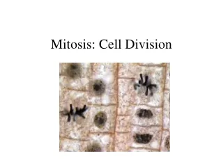
Cell division in eukaryotes • Chromosones consist of a DNA-protein complex called chromatin. Proteins include histones that aid in coiling of nucleic material in dense, visible chromatids • Mitosis duplicates chromosones to pairs of sister chromatids (duplication is very precise: only 1 error in 100,000) and sends each replication to opposite poles of the cell • Mitosis may be followed by cytokinesis
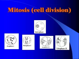
The cell cycle Interphase • 90% of time spent in Interphase • G1 = first growth phrase • S = duplication of DNA • G2 = second growth phase • 10% in dividing, or M phase • Mitosis - division of nucleus • Cytokinesis S-phase G1 G2 Mitosis M-phase
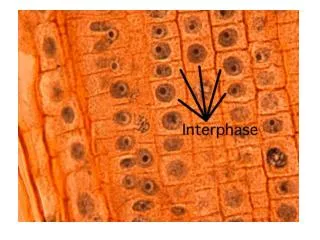
G2 Interphase • Characterized by • Well defined nucleus bound by nuclear envelope • Nucleoli • A pair of centrosomes adjacent to nucleus (division occurs in S-phase • (in animals) a pair of centrioles in each centrosome • (in animals) a microtubular array (aster) around each centrosome • Duplicated chromosones cannot be distinguished (chromatin too loosely packed)
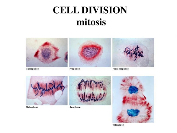
Prophase • Characterized by • Disappearance of nucleoli • Chromatin fibers condense into discrete chromosones composed of two identical sister chromatids joined at a centromere • Formation of mitotic spindle, composed of microtubules between the two centrosomes • Centrosomes move apart, migrating across surface of nuclear envelope
Prometaphase • Characterized by • Nuclear envelope fragments and dissolves • Spindle fibers extend from each pole towards the cell equator • Each chromatid has a specialized center called the kinetochore, located at the centromere • Kinetochore microtubules attach to the kinetochores • Non-kinetochore microtubules from each centrosome overlap each other
Metaphase • Characterized by • Centrosomes are at opposite poles of the cell • Chromosones migrate to the metaphase plate • Centromeres of all chromosones are aligned on metaphase plate • Kinetochores of sister chromatids face opposite poles, so that identical chromatids are attached to kinetochore fibers radiating from opposite ends of the parent cells • Entire structure formed by kinetochore and non-kinetochore fibers is termed the spindle
Anaphase • Characterized by • Sister chromatids split apart into separate chromosones, moving to opposite ends of the cell (depolymerization of microtubules at kinetochore end) • Chromosones move centromere-first, moving chromatids in a v-shape • Poles of cell move further apart, elongating cell At the end of anaphase, the two poles of the cell have identical collections of chromosones
Telophase • Characterized by • Non-kinetochore microtubules further elongate the cell • Daughter nuclei begin to form at the two poles • Nuclear envelope forms around chromosones from fragments of parent cell’s nuclear envelope • Nucleoli reappear • Chromatin protein uncoils and chromosones become less distinct
Cytokinesis • (in animal cells) Cleavage furrow forms and pinches off the two daughter cells • Done by a contractile ring of microtubules on cytoplasm side of membrane • (in plant cells) a cell plate (old metaphase plate) grows between the two daughter cells. • Cell membrane grows either side along length of cell plate, which becomes the cell wall By end of cytokinesis • Two separate daughter cells with genetically identical nuclei
Control of cell division • Cues include • Growth factors may bind with membrane receptors to promote division • Density dependent inhibition (affected by nutrients and space) • G1 phase has a ‘point of no return’, termed the restriction point, passed which a cell must go into the s-phase • If a cell doesn’t go past restriction point, goes into G0 phase - stasis • Cell size: ratio of cytoplasm to genome ration must be high enough to allow cell to go past restriction point
The mitotic clock • Still mostly unknown process: the onset of S-phase appears to commit cell to G2 and m-phase • Cell cycle events are synchronized by rhythmic changes in regulatory proteins called protein kinases that catalyze the phosphorylation of a target protein by ATP • Phosphorylation activates or inhibits the target protein’s activity • Cyclical changes in kinase activity are controlled by a second group of regulatory proteins called cyclins
Protein kinases that regulate cell cycles are cyclin-dependent kinases (Cdks) • Cdk concentration stay the same throughout the cell cycle. However, its activity varies • For example active MPF (maturation promoting factor) • Phosphorylates chromatin proteins, causing chromosones to condense in prophase • Phosphorylation of nuclear envelope in prometaphase promotes fragmentation
Interphase Interphase mitosis mitosis mitosis • Cyclin, produced throughout the cell cycle, accululates during interphase • Cyclin combines with cdk to make active MPF, which peaks in concentration with the peak in cyclin • MPF initiates mitosis • At the end of mitosis, MPF activates an enzyme that destroys cyclin Relative concentration time
Cancers - cells lacking division control • Cancerous cells do not respond to standard cellular controls, growing and dividing until nutrients are exhausted • Cells that become cancerous are said to have transformed • Immune system usually destroys such cells, but if not, cancerous cells coalesce to tumors • If cells remain at the tumor, then the tumor is benign • If cells spread out, then tumor is malignant - spread is called metastasis • Cause of cancerous cells is probably caused by alteration of genes that control cell division