Download
1 / 100
1k likes | 1.05k Views
Explore the architecture and functions of the central and peripheral nervous systems, spinal cord anatomy, and spinal reflexes.

E N D
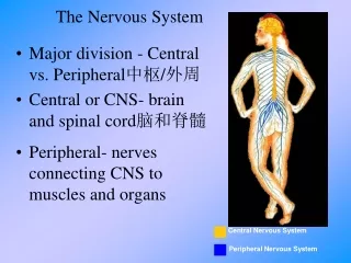
Central Nervous System Peripheral Nervous System The Nervous System • Major division - Central vs. Peripheral中枢/外周 • Central or CNS- brain and spinal cord脑和脊髓 • Peripheral- nerves connecting CNS to muscles and organs
中枢/外周神经功能分类. ♠ Sensory division brings information regarding the int. or ext. environment into the CNS ♠ Motor division issues directives out to muscle or glandular tissue ♣Somatic nervous system services skeletal muscle ♣ Autonomic nervous system services smooth &cardiac muscle: ♦ Sympathetic nervous system is responsible for the "fight-or-flight"response; ♦ Parasympathetic nervous system is responsible for the "rest-anddigest"response
Divisions of the Nervous System • Central Nervous System • Peripheral Nervous System • nerves • cranial nerves • spinal nerves
The Spinal Cord 1. External features: • Location: foramen magnum -- lower border of first lumbar vertebra • Shape: A long cylindrical structure • Enlargements: cervical enlargement lumbar [lumbosacral] enlargement • Conus medullaris脊髓圆锥 • Filum terminale : a condensation ofpia mater, S2,终丝
Conus edullaris :caudal to the lumbosacral enlargement the spinal cord tapers gradully& becomes the conical termination Filum termination: a condensation of pia mater forms~ Cauda equina: 马尾lumbosacral roots descend ,sourrounding the filum terminale..
sulcus • main longitudinal fissure and sulci on surface: Cauda equina posterior lateral posterior median sulcus anterior lateral anterior median Filum terminale sulcus fissure
Spinal segment: It's a part of spinal cord, which is connected with the rootlets of a pair of spinal nerve. • 31 segments 8 cervical segments 12 thoracic segments 5 lumbar segments 5 sacral segments 1 coccygeal segments
Corresponding relationship between spinal segments and vertebrae spinal segments correspond to vertebrae C1-C4 C1-C4 C5 ~ T8, T l~ T4 C4 - C7, C7 ~ T3 T5 ~ T8 T3 ~ T6 T9-T12 T6-T9 L1-L5 T10-T12 S l~S5,Co1 LI
posterior horn central canal 2. Internal structure • The central canal • Gray matter: • parts: • Lateral horn (only extends from Tl to L3 segments.) • gray commissure (anterior and posterior ) Intermediate zone Lateral horn anterior horn Gray matter gray commissure
Gray matter anterior horn posterior horn intermediate zone White matter anterior funiculus posterior funiculus Lateral funiculus
posterior funiculus posterior median sulcus posterior lateral sulcus • White matter: • parts: lateral funiculus anterior lateral sulcus anterior funiculus anterior white commissure anterior median fissure
Main tracts (or fasciculi):long ascending tracts: • fasciculus gracilis薄束: come from sacral, lumbar and lower eight thoracic nerves, terminate upon nucleus gracilis • fascicules cuneatus楔束: come from cervical and upper fouth thoracic nerves, terminate upon nucleus cuneatus. • *conduct the fine [discriminating] tactile (e.g.two-point discrimination ) & kinesthetic sense (e.g.position , movement &vibration)of the ipsilateral trunk & limbs. fascicules cuneatus fascicules gracilis
thalamus • lateral & anterior spinothalamic tracts: 脊髓丘脑束 • *in corresponding funiculus • *arises from opposite nucleus proprius • *terminate on thalamus • *conduct pain, thermal and rough tactile sense of opposite trunk and limbs anterior spinothalamic tracts anterior spinothalamic tracts nucleus proprius
main long descending tracts lateral corticospinal tract: 皮质脊髓侧束 *arises from opposite cerebral cortex *descends through lateral funiculus of spinal cord *terminate on ipsilateral anterior horn (lateral group) *controls the contraction of skeletal muscles of ipsilateral limbs anterior corticospinal tract: *controls the movement of bilateral muscles of trunk lateral corticospinal tract anterior corticospinal tract
Functions: • To convey afferent impulses, which come from somatic and visceral receptors to the brain, and conduct efferent impulses from brain to effectors. • Related to reflexes,e.g., the stretch reflex, the reflex of bladder & rectal emptying
Spinal Reflexes • Programmed stereotypical reactions that occur in response to stimuli • Simplest reflex: monosynaptic stretch reflex • Reflexes are subject to higher level control
P e r i p h e r a l N e r v o u s S y s t e m S k e l e t a l A u t o n o m i C内脏 ( S o m a t i c )躯体 S y m p a t h e t i C交感 P a r a s y m p a t h e t i C副交感 Peripheral Nervous System
Brain Sensory Neuron Motor Neuron Skin receptors Interneuron Muscle Somatic System • Nerves to/from spinal cord • control muscle movements • somatosensory inputs • Both Voluntary and reflex movements • Skeletal Reflexes • simplest is spinal reflex arc
Autonomic System • Two divisions: • Sympathetic nerve交感神经 • Parasympatheitic nerve副交感神经 • Control involuntary functions • heartbeat • blood pressure • respiration • perspiration • digestion • Can be influenced by thought and emotion
CENTRAL NERVOUS SYSTEM SYMPATHETIC Brain Dilates pupil Stimulates salivation Salivary glands Relaxes bronchi Spinal cord Lungs Accelerates heartbeat Heart Inhibits activity Stomach Pancreas Stimulates glucose Liver Adrenal gland Secretion of adrenaline, nonadrenaline Kidney Relaxes bladder Sympathetic ganglia Stimulates ejaculation in male Sympathetic • “ Fight or flight” response • Release adrenaline and noradrenaline • Increases heart rate and blood pressure • Increases blood flow to skeletal muscles • Inhibits digestive functions
CENTRAL NERVOUS SYSTEM PARASYMPATHETIC Brain Contracts pupil Stimulates salivation Constricts bronchi Spinal cord Slows heartbeat Stimulates activity Stimulates gallbladder Gallbladder Contracts bladder Stimulates erection of sex organs Parasympathetic • “ Rest and digest ” system • Calms body to conserve and maintain energy • Lowers heartbeat, breathing rate, blood pressure
Autonomic nervous system controls physiological arousal Sympathetic division (arousing) Parasympathetic division (calming) Pupils dilate EYES Pupils contract Decreases SALVATIONIncreases Perspires SKIN Dries Increases RESPERATIONDecreases Accelerates HEARTSlows Inhibits DIGESTIONActivates Secrete stress hormones ADRENAL GLANDS Decrease secretion of stress hormones Summary of autonomic differences
Sympathetic Division • Myelinated preganglionic exit spinal cord in ventral roots at T1 to L2[L3] levels. • Travel to ganglia at different levels to synapse with postganglionic neurons. • Divergence: • Preganglionic fibers branch to synapse with numerous postganglionic neurons.
Sympathetic Division • Axons of postganglionic neurons are unmyelinated to the effector organ. • Preganglionic neuron is short. • Post-ganglionic neuron is long.
Parasympathetic Division • Preganglionic fibers originate in midbrain, medulla, and pons; and in the 2-4 sacral levels of the spinal cord. • Preganglionic fibers synapse in ganglia located next to or within organs innervated. • Do not travel within spinal nerves. • Do not innervate blood vessels, sweat glands,and arrector pili muscles.
Parasympathetic Division • 4 of 12 pairs of cranial nerves contain preganglionic parasympathetic fibers. • Preganglionic fibers are long, postganglionic fibers are short. • Vagus: • Innervate heart, lungs esophagus, stomach, pancreas, liver, small intestine and upper half of the large intestine.
Parasympathetic Division • Preganglionic fibers from the sacral level innervate the lower half of large intestine, the rectum, urinary and reproductive systems.
THE PERIPHERAL NERVOUS SYSTEM ---- The Spinal Nerves 1. General Description • 31 Pairs of Spinal Nerves Cervical Nerves : 8 pairs Thoracic Nerves : 12 pairs Lumbar Nerves : 5 pairs Sacral Nerves : 5 pairs Coccygeal Nerves : 1 pair
Four Types of Fibers in Spinal Nerves • The Somatic afferent (sensory) Fibers - The Visceral afferent (sensory) Fibers • The Somatic efferent (motor) Fibers - The Visceral efferent (motor) Fibers
Four Branches of a Spinal Nerves • Four Branches of a Spinal Nerves • Anterior BranchPosterior Branch • Meningeal Branch Communicating Branches
Four Plexuses • Cervical Plexus颈丛 • Brachial Plexus臂丛 • Lumbar Plexus腰丛 • Sacral Plexus骶丛 • The thoracic nerves are separated from each other.
The cervical Plexus • The Formation of the Cervical Plexus: • The anterior branches of the 1st to 4th cervical nerves [C1~4]
The Superficial Branches(sternocleidomastoid m.) The lesser occipital nerve The great auricular nerve The transverse nerve of neck The supraclavicular nerves
The Deep Branches • The phrenic nerve
The Brachial Plexus (scalene fissure, axilla) • formation:root(C5 – T1) trunk division cord
branches and distribution Brachial plexus : the long thoracic the musculocutaneous nerve lateral cord lateral pectoral nerve the median nerve Medial cord the ulnar nerve medial pectoral nerve the radial nerve Posterior cord the axillary nerve the thoracodorsal nerve
the musculocutaneous n. the axillary n. the long thoracic n. medial pectoral n. lateral pectoral n. median n. the ulnar n. the radial n. the thoracodorsal n.
The musculocutaneous nerve ♠ Major end branch of lateral cord ♠ Runs down lateral aspect of upper limb ♠ Innervates forearm flexors (biceps, brachialis) and provides cutaneous sensation for lateral forearm • The median nerve ♠ Does not branch in arm (humeral region of upper limb) ♠ In forearm, innervates flexors and skin ♠ In hand, innervates some muscles of lateral palm ♠ The median nerve serves muscles which pronate the forearm,flex the wrist and fingers and oppose the thumb
The ulnar nerve ♠ Branches off median cord. ♠ Runs along medial aspect of arm, swings behind the medial epicondyle, then parallels the ulna as it courses down the medial forearm ♠ Innervates flexor carpi ulnaris & flexor digitorum profundus;Supplies most of the muscles & skin of the medial hand ♠ Along with median nerve, the ulnar nerve produces wrist &finger flexion &adduction & abduction of medial fingers ♣ The "funny bone" is the ulnar nerve as it passes behind the medial epicondyle. ♠ Loss of function of the ulnar nerve results in clawhandas the little and ring fingers become hyperextended at the knuckles and flexed at the distal interphalangeal joints
The radial nerve Largest branch of the brachial plexus; continuation of the posterior cord,wraps around posterior humerus, swings up & over the lateral epicondyle &the divides into a superficial branch &a deep branch ♣ Superficial branch,runs along lateral edge of radius ♣ Deep branch,runs into posterior forearm ♠ Radial nerve innervates nearly all extensors in the upper limb,produces extension of elbow (triceps), supination of forearm (supinator, brachioradialis, but not biceps), extension of wrist & fingers (forearm and digital extensors), and abduction of thumb ♠ Loss of function of the radial nerve results in wrist drop, an inability to extend the hand at the wrist


