Download
1 / 18
180 likes | 185 Views
Understand the structure and function of DNA, the molecule that carries genetic information and controls cellular processes. Learn about DNA replication and its role in determining traits.

E N D
DNA • Within the nucleus of almost all of your cells 46 DNA molecules or chromosomes contain approx. 20-25000 genes. • These genes act as a blueprint directing which proteins are to be synthesised by the cell. • By controlling cell functioning, DNA is able to determine the characteristics of a person. • DNA is unique among molecules because its structure allows it to manufacture another molecule, identical to itself, and in doing so transmit its genetic code from parents to their children and from one cell to another as new cells are formed.
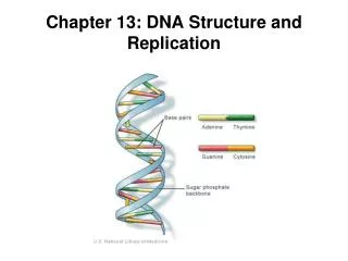
Structure of DNA • DNA is a condensation polymer constructed from four monomers known as nucleotides. • Each nucleotide consists of a phosphate group, a deoxyribose sugar group and one of the four bases. • The four bases are adenine (A), cytosine (C), thymine (T) and Guanine (G). • Adenine and Guanine are derivatives of the organic compound purine and as a results are referred to as purine bases. • Cytosine and thymine are derivatives of another compound, pyrimidine, and are referred to as pyrimidine bases. • The base forms a covalent bond to the carbon atom labelled C1 in the deoxyribose molecule. • The phosphate group bonds to the carbon atom labelled C5. • A water molecule is produced when each bond is formed.
* Indicates the site where the base bonds to the C1 deoxyribose carbon atom.
A simplified presentation of the condensation reaction between nucleotides to form a section of DNA.
Primary Structure of the Polymer • Covalent bonds are responsible for producing the primary structure of DNA. • A covalent bond is formed between the 5’-phosphate group on one nucleotide and the 3’-hydroxy on the deoxyribose of another. • One end of the DNA strand has a hydroxyl group and is called the 3’ end. • The end of the polymer with the phosphate group is called the 5’ end. • In this way, nucleotides undergo condensation polymerisation to form a long chain of nucleotides. • A base is also covalently bonded to the sugar group at the C1 site via a condensation reaction. • It is the sequence of bases along the sugar-phopshate backbone that forms the primary structure and is the basis for the genetic code.
Secondary Structure of the Polymer • As is the case in proteins, hydrogen bonding is responsible for maintaining the secondary structure of DNA. • For a given species, the percentage of each of the four bases is the same in all cells and is characteristic of the organism. • The mole amounts of thymine (a purine) and adenine (a pyrimidine) are equal. • The mole amounts of cytosine (a pyrimidine) and guanine (a purine) are equal.
Secondary Structure of the Polymer cont… • By considering the structures of the base pair A and T, it can be seen that they are able to fit together. • It is possible for two hydrogen bonds to form between the dipole negative on the nitrogen and oxygen atoms and the dipole positive on hydrogen atoms on the adjacent base. • With base pairs C and G three hydrogen bonds can form between the nitrogen and oxygen atoms of one base and the hydrogen atoms on the adjacent base.
Secondary Structure of the Polymer cont… • The secondary structure of DNA is a pair of DNA polynucleotide strands held together by hydrogen bonding between the bases A-T and C-G. • The structure twists around to form a right-handed double helix. • The sugar-phosphate backbone forms the handrails and the pairs of bases the steps. • The pairing of bases is referred to as complementary base pairing. • The DNA strands align in opposite direction.
Tertiary Structure of the Polymer • The phosphate groups in the backbone of the double helix give DNA molecules a negative charge, and this enables the molecules to interact with a group of proteins called histones. • The DNA molecules wrap around histones and become super coiled. • This allows the DNA to be very tightly and efficiently packaged, each molecule forming a structure known as a chromosome.
Replication • The division of plant and animal cells involves a mechanism that generates an exact copy of their DNA. • The DNA double helix partially unwinds, as hydrogen bonds between the two DNA strands are broken. • Enzymes catalyse this process. • The bases exposed on the separated strands then act as a template to which new nucleotides attach by hydrogen bonds between complementary base pair, C and G, A and T. • These bases then undergo a condensation polymerisation reaction catalysed by the enzyme DNA polymerase to form two exact copies of the original DNA double helix.
How does DNA control biochemical processes? • Alternative sequences of nucleotides can occur at specific positions on a given chromosome. • The alternative sequences are referred to as alleles. • Alleles give rise to most of the variation we see between people. • Triple code – a sequence of three DNA bases codes for an amino acid. • RNA decodes the information on DNA in the nucleus of the cell where protein synthesis takes place. • Another type of RNA controls the selection and sequencing of the amino acids that form proteins. Read Sickle-Cell Anaemia and Cystic Fibrosis
Forensic Applications - Electrophoresis • Electrophoresis is an analytical technique, similar to chromatography, that allows large charged molecules, DNA and proteins, to be separated and quantitively determined. • In electrophoresisthe sample solution is placed into a “well” in a block of conducting electrolytic gel, sitting between (+) and (-) electrodes.
Forensic Applications – Electrophoresis cont… • negative ions, such as DNA fragments, in the sample move toward the positive electrode. • The rate of travel depends on their size, how many base pairs there are. • for DNA analysis, the sample “well” is placed near the negative electrode, as DNA fragments, with their HPO4- phosphate linkages, are always negatively charged. • the distance the fragments move is compared against the distance travelled by known standards. • the separated components can be made visible by adding fluorescent dyes or radioactive markers.
Forensic Applications – DNA Profiling • The sections of DNA that do not code for the production of proteins, non-coding regions are more useful for forensic scientists because they differ significantly from person to person • Of particular interest is the number of times a particular sequence of bases may be repeated eg. on chromosome 5 the sequence AGAT is repeated between 7 and 15 times, depending on the person. • When you examine 10 different chromosome sites, counting the number of base sequence repeats, an accurate DNA profile identification is assured.
Forensic Applications – Polymerase Chain Reaction (PCR) • a major problem with early DNA forensics was obtaining a big enough biological sample, blood, skin, semen etc. to analyse. • PCR has enabled forensic scientists to produce a DNA profile from an extremely small sample • PCR utilizes DNA’s ability to replicate itself, making multiple copies of the sections of the DNA of interest
Forensic Applications - PCR • PCR involves 3 steps … 1. Denaturation … heating breaks the H-bonding between DNA strands 2. Annealing … the sample is cooled and primers are added to bond to the start and end of the base sequence to be copied 3. Elongation … the sample is warmed and polymerase enzyme catalyst assists the addition of complementary base pairs and the formation of H-bonds between the DNA strands • The amplified DNA fragments are then separated using gel electrophoresis and the analysis of the number of base sequence repeats for 10 different chromosome locations performed automatically.