Download
1 / 1
10 likes | 158 Views
The Comparison of Three Dimensional Brain Vessel Image according To Scan Methods Department of Radiology, Yonsei University Health System Kyoo Hyun Kim, hong sik Kim, Ho Yung Hwang, Sung Ho Kang, Jae Sik Lim. Figure 3. Dual Energy scan method (function tool). A B.

E N D
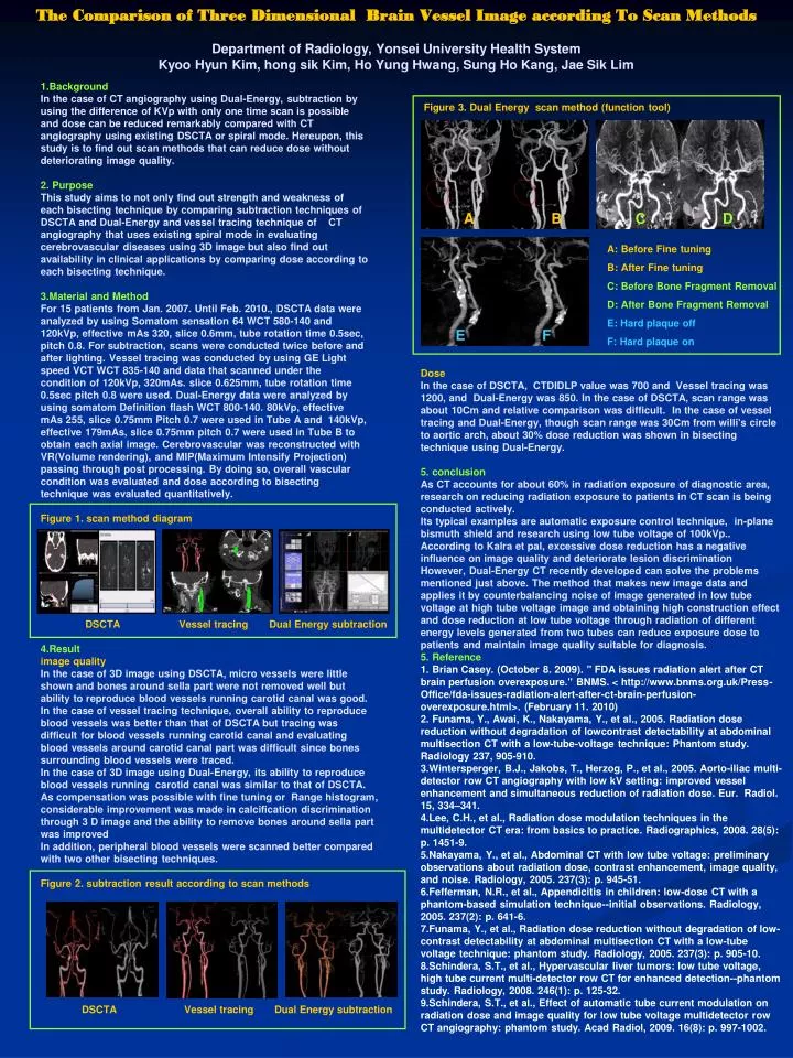
The Comparison of Three Dimensional Brain Vessel Image according To Scan Methods Department of Radiology, Yonsei University Health System Kyoo Hyun Kim, hong sik Kim, Ho Yung Hwang, Sung Ho Kang, Jae Sik Lim Figure 3. Dual Energy scan method (function tool) A B C D A: Before Fine tuning B: After Fine tuning C: Before Bone Fragment Removal D: After Bone Fragment Removal E: Hard plaque off F: Hard plaque on E F DSCTA Vessel tracing Dual Energy subtraction 1.Background In the case of CT angiography using Dual-Energy, subtraction by using the difference of KVp with only one time scan is possible and dose can be reduced remarkably compared with CT angiography using existing DSCTA or spiral mode. Hereupon, this study is to find out scan methods that can reduce dose without deteriorating image quality. 2. Purpose This study aims to not only find out strength and weakness of each bisecting technique by comparing subtraction techniques of DSCTA and Dual-Energy and vessel tracing technique of CT angiography that uses existing spiral mode in evaluating cerebrovascular diseases using 3D image but also find out availability in clinical applications by comparing dose according to each bisecting technique. 3.Material and Method For 15 patients from Jan. 2007. Until Feb. 2010., DSCTA data were analyzed by using Somatom sensation 64 WCT 580-140 and 120kVp, effective mAs 320, slice 0.6mm, tube rotation time 0.5sec, pitch 0.8. For subtraction, scans were conducted twice before and after lighting. Vessel tracing was conducted by using GE Light speed VCT WCT 835-140 and data that scanned under the condition of 120kVp, 320mAs. slice 0.625mm, tube rotation time 0.5sec pitch 0.8 were used. Dual-Energy data were analyzed by using somatom Definition flash WCT 800-140. 80kVp, effective mAs 255, slice 0.75mm Pitch 0.7 were used in Tube A and 140kVp, effective 179mAs, slice 0.75mm pitch 0.7 were used in Tube B to obtain each axial image. Cerebrovascular was reconstructed with VR(Volume rendering), and MIP(Maximum Intensify Projection) passing through post processing. By doing so, overall vascular condition was evaluated and dose according to bisecting technique was evaluated quantitatively. Figure 1. scan method diagram Dose In the case of DSCTA, CTDIDLP value was 700 and Vessel tracing was 1200, and Dual-Energy was 850. In the case of DSCTA, scan range was about 10Cm and relative comparison was difficult. In the case of vessel tracing and Dual-Energy, though scan range was 30Cm from willi's circle to aortic arch, about 30% dose reduction was shown in bisecting technique using Dual-Energy. 5. conclusion As CT accounts for about 60% in radiation exposure of diagnostic area, research on reducing radiation exposure to patients in CT scan is being conducted actively. Its typical examples are automatic exposure control technique, in-plane bismuth shield and research using low tube voltage of 100kVp.. According to Kalra et pal, excessive dose reduction has a negative influence on image quality and deteriorate lesion discrimination However, Dual-Energy CT recently developed can solve the problems mentioned just above. The method that makes new image data and applies it by counterbalancing noise of image generated in low tube voltage at high tube voltage image and obtaining high construction effect and dose reduction at low tube voltage through radiation of different energy levels generated from two tubes can reduce exposure dose to patients and maintain image quality suitable for diagnosis. 5. Reference 1. Brian Casey. (October 8. 2009). " FDA issues radiation alert after CT brain perfusion overexposure." BNMS. < http://www.bnms.org.uk/Press-Office/fda-issues-radiation-alert-after-ct-brain-perfusion-overexposure.html>. (February 11. 2010) 2. Funama, Y., Awai, K., Nakayama, Y., et al., 2005. Radiation dose reduction without degradation of lowcontrast detectability at abdominal multisection CT with a low-tube-voltage technique: Phantom study. Radiology 237, 905-910. 3.Wintersperger, B.J., Jakobs, T., Herzog, P., et al., 2005. Aorto-iliac multi-detector row CT angiography with low kV setting: improved vessel enhancement and simultaneous reduction of radiation dose. Eur. Radiol. 15, 334–341. 4.Lee, C.H., et al., Radiation dose modulation techniques in the multidetector CT era: from basics to practice. Radiographics, 2008. 28(5): p. 1451-9. 5.Nakayama, Y., et al., Abdominal CT with low tube voltage: preliminary observations about radiation dose, contrast enhancement, image quality, and noise. Radiology, 2005. 237(3): p. 945-51. 6.Fefferman, N.R., et al., Appendicitis in children: low-dose CT with a phantom-based simulation technique--initial observations. Radiology, 2005. 237(2): p. 641-6. 7.Funama, Y., et al., Radiation dose reduction without degradation of low-contrast detectability at abdominal multisection CT with a low-tube voltage technique: phantom study. Radiology, 2005. 237(3): p. 905-10. 8.Schindera, S.T., et al., Hypervascular liver tumors: low tube voltage, high tube current multi-detector row CT for enhanced detection--phantom study. Radiology, 2008. 246(1): p. 125-32. 9.Schindera, S.T., et al., Effect of automatic tube current modulation on radiation dose and image quality for low tube voltage multidetector row CT angiography: phantom study. Acad Radiol, 2009. 16(8): p. 997-1002. DSCTA Vessel tracing Dual Energy subtraction 4.Result image quality In the case of 3D image using DSCTA, micro vessels were little shown and bones around sella part were not removed well but ability to reproduce blood vessels running carotid canal was good. In the case of vessel tracing technique, overall ability to reproduce blood vessels was better than that of DSCTA but tracing was difficult for blood vessels running carotid canal and evaluating blood vessels around carotid canal part was difficult since bones surrounding blood vessels were traced. In the case of 3D image using Dual-Energy, its ability to reproduce blood vessels running carotid canal was similar to that of DSCTA. As compensation was possible with fine tuning or Range histogram, considerable improvement was made in calcification discrimination through 3 D image and the ability to remove bones around sella part was improved In addition, peripheral blood vessels were scanned better compared with two other bisecting techniques. Figure 2. subtraction result according to scan methods