Download
1 / 119
1.19k likes | 1.3k Views
What is the leading cause of death in the U.S.? (2005). Heart disease: 652,091 Cancer: 559,312 Stroke (cerebrovascular diseases): 143,579 Chronic lower respiratory diseases: 130,933 Accidents (unintentional injuries): 117,809 Diabetes: 75,119 Alzheimer's disease: 71,599

E N D
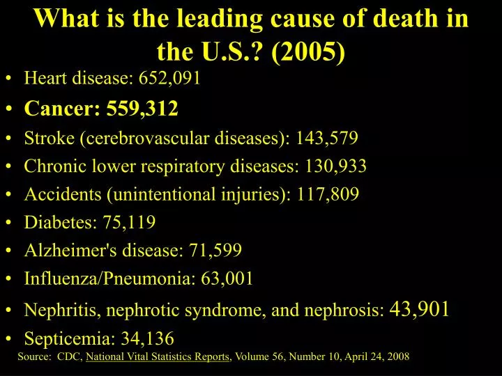
What is the leading cause of death in the U.S.? (2005) • Heart disease: 652,091 • Cancer: 559,312 • Stroke (cerebrovascular diseases): 143,579 • Chronic lower respiratory diseases: 130,933 • Accidents (unintentional injuries): 117,809 • Diabetes: 75,119 • Alzheimer's disease: 71,599 • Influenza/Pneumonia: 63,001 • Nephritis, nephrotic syndrome, and nephrosis: 43,901 • Septicemia: 34,136 Source: CDC, National Vital Statistics Reports, Volume 56, Number 10, April 24, 2008
Leading Causes of U. S. Deaths (2006) • Heart disease: 631,636 • Cancer: 559,888 • Stroke (cerebrovascular diseases): 137,119 • Chronic lower respiratory diseases: 124,583 • Accidents (unintentional injuries): 121,599 • Diabetes: 72,449 • Alzheimer's disease: 72,432 • Influenza and Pneumonia: 56,326 • Nephritis, nephrotic syndrome, and nephrosis: 45,344 • Septicemia: 34,234 Source: CDC, National Vital Statistics Reports, Volume 57, Number 14, April 17, 2009
The Cell CycleChapter 12- A.P. Biology Mr. Knowles Liberty Senior High School
Figure 12.1 • Overview: The Key Roles of Cell Division • The continuity of life: • is based upon the reproduction of cells, or cell division
100 µm (a) Reproduction. An amoeba, a single-celled eukaryote, is dividing into two cells. Each new cell will be an individual organism (LM). Figure 12.2 A Unicellular organisms: • Reproduce by cell division.
Origin of replication Cell wall Plasma Membrane E. coli cell Bacterial Chromosome Chromosome replication begins. Soon thereafter, one copy of the origin moves rapidly toward the other end of the cell. Two copies of origin 1 Replication continues. One copy ofthe origin is now at each end of the cell. 2 Origin Origin Replication finishes. The plasma membrane grows inward, and new cell wall is deposited. 3 Two daughter cells result. 4 • In binary fission: • The bacterial chromosome replicates • The two daughter chromosomes actively move apart Figure 12.11
Cell Replication in Bacteria • Cell division in bacteria is controlled by the size of the cell; volume of the cytoplasm. • Bacteria replicate by binary fission. • >22 enzymes copy the DNA as a circle. • Both copies of the DNA are attached to the plasma membrane.
The Evolution of Mitosis • Since prokaryotes preceded eukaryotes by billions of years: • It is likely that mitosis evolved from bacterial cell division • Certain protists: • Exhibit types of cell division that seem intermediate between binary fission and mitosis carried out by most eukaryotic cells
(a) Prokaryotes. During binary fission, the origins of the daughter chromosomes move to opposite ends of the cell. The mechanism is not fully understood, but proteins may anchor the daughter chromosomes to specific sites on the plasma membrane. Bacterial chromosome Chromosomes (b) Dinoflagellates. In unicellular protists called dinoflagellates, the nuclear envelope remains intact during cell division, and the chromosomes attach to the nuclear envelope. Microtubules pass through the nucleus inside cytoplasmic tunnels, reinforcing the spatial orientation of the nucleus, which then divides in a fission process reminiscent of bacterial division. Microtubules Intact nuclear envelope (c) Diatoms. In another group of unicellular protists, the diatoms, the nuclear envelope also remains intact during cell division. But in these organisms, the microtubules form a spindle within the nucleus. Microtubules separate the chromosomes, and the nucleus splits into two daughter nuclei. Kinetochore microtubules Intact nuclear envelope Kinetochore microtubules (d) Most eukaryotes. In most other eukaryotes, including plants and animals, the spindle forms outside the nucleus, and the nuclear envelope breaks down during mitosis. Microtubules separate the chromosomes, and the nuclear envelope then re-forms. Centrosome Fragments of nuclear envelope A hypothetical sequence for the evolution of mitosis Figure 12.12 A-D
200 µm 20 µm (c) Tissue renewal. These dividing bone marrow cells (arrow) will give rise to new blood cells (LM). (b) Growth and development. This micrograph shows a sand dollar embryo shortly after the fertilized egg divided, forming two cells (LM). Figure 12.2 B, C Multicellular organisms depend on cell division for: • Development from a fertilized cell • Growth • Repair
Cell Division in Eukaryotic Cells • Eukaryotic cells are much larger. • Also have larger genomes (the sum of an organisms’s genetic information). • Eukaryotic DNA is organized into much more complex structures. • Why?
Figure 12.3 50 µm The DNA molecules in a cell • Are packaged into chromosomes.
Chromosomes of Eukaryotes • Chromosomes are composed of chromatin - a DNA/protein complex. • Chromatin = 40% DNA + 60% Protein. • Every 200 nucleotides , the DNA duplex coils around a core of eight histoneproteins = Nucleosome.
Regions of Chromosomes are Not the Same • Condensed portions of chromatin - Heterochromatin - are not being expressed; genes are turned off. May never be turned on. • Other portions of chromosome are decondensed and are being actively transcribed - Euchromatin. These areas are only condensed during cell division.
Chromosome Terminology • The two copies of each chromosome in somatic cells - homologous chromosomes (homologues). Are these the same? • Before cell division, each homologue replicates --> two sister chromatids that are joined at the centromere.
How Many Chromosomes are in Cells? • Most body or somatic cells have two copies of almost identical chromosomes - diploid. Ex. Humans have 23 types of chromosomes X 2 = 46 (2n or diploid number). • Sex cells or gametes (egg and sperm) have only one copy of each chromosome - haploid. Ex. Human sperm/egg = 23 chromosomes.
How Many Chromosomes are in Cells? • Some somatic cells are truly unusual. • Human liver cells have 4 copies of all chromosomes - tetraploid. • Other cells RBC’s have no nucleus and therefore, no chromosomes.
INTERPHASE S(DNA synthesis) G1 CytokinesisMitosis G2 MITOTIC(M) PHASE Figure 12.5 Phases of the Cell Cycle • The cell cycle consists of • The mitotic phase • Interphase G0
Telomeres and Human Disease Telomere Length in Human Sperm and Aging Video: Scientific American Frontiers: Never Say Die, VT 571.879 SCI
0.5 µm A eukaryotic cell has multiplechromosomes, one of which is represented here. Before duplication, each chromosomehas a single DNA molecule. Chromosomeduplication(including DNA synthesis) Once duplicated, a chromosomeconsists of two sister chromatidsconnected at the centromere. Eachchromatid contains a copy of the DNA molecule. Centromere Sisterchromatids Separation of sister chromatids Mechanical processes separate the sister chromatids into two chromosomes and distribute them to two daughter cells. Centromeres Sister chromatids Figure 12.4 • Each duplicated chromosome: • Has two sister chromatids, which separate during cell division
PROMETAPHASE G2 OF INTERPHASE PROPHASE Centrosomes(with centriole pairs) Aster Fragmentsof nuclearenvelope Early mitoticspindle Kinetochore Chromatin(duplicated) Centromere Nonkinetochoremicrotubules Kinetochore microtubule Nucleolus Chromosome, consistingof two sister chromatids Nuclearenvelope Plasmamembrane Figure 12.6 • Mitosis consists of five distinct phases • Prophase • Prometaphase
METAPHASE ANAPHASE TELOPHASE AND CYTOKINESIS Metaphaseplate Cleavagefurrow Nucleolusforming Nuclear envelopeforming Daughter chromosomes Centrosome at one spindle pole Spindle Figure 12.6 • Metaphase • Anaphase • Telophase
Kinetochores are proteins attached to the centrosome. They are used to anchor the spindle fibers to the chromosome.
The Mitotic Spindle: A Closer Look • The mitotic spindle: • Is an apparatus of microtubules that controls chromosome movement during mitosis.
Aster Centrosome MetaphasePlate Sisterchromatids Kinetochores Overlappingnonkinetochoremicrotubules Kinetochores microtubules 0.5 µm Microtubules Chromosomes Figure 12.7 Centrosome 1 µm • Some spindle microtubules: • Attach to the kinetochores of chromosomes and move the chromosomes to the metaphase plate
EXPERIMENT 1 The microtubules of a cell in early anaphase were labeled with a fluorescent dye that glows in the microscope (yellow). Kinetochore Spindlepole Figure 12.8 • In anaphase, sister chromatids separate: • And move along the kinetochore microtubules toward opposite ends of the cell
Nonkinetechore microtubules from opposite poles • Overlap and push against each other, elongating the cell • In telophase: • Genetically identical daughter nuclei form at opposite ends of the cell