Download
1 / 42
420 likes | 641 Views
Virus Life Cycles in 3D. The Art of Reconstruction. Virus Life Cycle. In order to survive, viruses must be able to do the following: 1. Find a host cell it can replicate in 2. Bind to that cell 3. Enter the cell 4. Release its genome in order to replicate 5. Replicate its genome

E N D
Virus Life Cycles in 3D The Art of Reconstruction
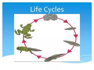
Virus Life Cycle • In order to survive, viruses must be able to do the following: • 1. Find a host cell it can replicate in • 2. Bind to that cell • 3. Enter the cell • 4. Release its genome in order to replicate • 5. Replicate its genome • 6. Transcribe and translate its viral proteins • 7. Package its genome and proteins • 8. Escape from the cell
Virus Life Cycle • All these processes can be visualized by cryo • These visualizations allows for a better understanding of viruses and may lead to vaccination development • For each virus, there is a unique life cycle but all viruses accomplish the same steps in order to survive
Enveloped Virus • Semliki Forest Virus is an enveloped Alphavirus • It has 2 transmembrane proteins (E1 and E2) in its envelope • The virus binds to the cellular receptor, endocytosed, and fuses with the endosome membrane to release its nucleocapsid for replication
Non enveloped Viruses • Poliovirus is a non enveloped virus in the Picornavirus family • It differs from SFV in that when it binds to its cellular receptor, it goes through a conformational change. • This conformational change may facilitate the release of genome into the cell for replication • Also releases from the cell by lysis instead of budding
Cell Attachment • The first step in viral replication is to be able to bind to the correct host cell. • Virus recognize host cells by certain receptors. • Bind to these receptors through specific interactions. • Binding sites on viruses are typically conserved to ensure survival
The Receptor binding region of HRV14 • Picornaviruses shield their receptor binding site in a region called the canyon in order to protect it from antibodies. • Must be conserved so that the virus can bind to the correct cell in order to replicate. • HRV16 + ICAM-1 interaction was one of the first to be studied through cryo • Was believed that the binding site for ICAM-1 was located in the canyon region of HRV16
HRV16 complexed with ICAM-1 • HRV16 complexed with the 2 N terminal domains of ICAM-1 • The footprint of ICAM-1 was centered over the canyon as predicted showing that the canyon was in fact the binding site of the receptor
Simian Rotavirus • VP4 of rotavirus is important to the viral life cycle • It is a determinant of virulence, has hemagglutination activity and is also a neutralization site • The reconstruction showed that VP4 extends from the surface of the virus, which may then be able to bind to the cellular receptor more easily
Activation • Viruses must be stable enough to survive the extracellular environment but must also be unstable enough to release their genome when they reach susceptible cells. • Certain conformational changes must occur in the virus when it reaches the proper environment in order to release its genome in the correct place and at the correct time.
SFV Spikes • SFV has a spike protruding from its envelope comprised of E1, E2, and E3 • Reconstruction showed that the spike has a hole in its center • From previous studies, E3 was determined to be on the outside of the spike • Preferential extraction and reconstruction comparison determined that E1made up the outside of the spike while E2 extended from the center
SFV spike conformational changes • In order to determine the conformational changes needed for activation, the particles were treated with low pH and vitrified within milliseconds • Comparison between treated and untreated particle reconstructions showed that E1 and E2 move around each other • E2 is the receptor binding portion while E1 is the membrane fusion protein • E2 moves outward while E1 moves inward to form a trimmer and trigger fusion
Adenovirus • Adenovirus is made up of hexons and two proteins at the five fold vertice: penton base and fiber • It binds two receptors: CAR and an integrin • CAR binds to the fiber while the penton base binds the integrin and causes activation
Adenovirus uses 2 receptors CAR Integrins
Structural changes in penton • The conformational changes needed for activation were determined by comparing particles which had the fiber attached and which did not • A small region which was determined to contain the RGD sequence by MAb binding changed orientation
Genome Release • The genome of the virus is released in order to make viral proteins and reproduce the genome. • Viruses can employ several strategies to do this: injection, release into the cytoplasm, release into the nucleus • Exception: Reoviruses
Flock House Virus • FHV is comprised of 180 copies of a single protein which undergoes a post assembly cleavage • The cleavage produces γ peptides which lie in different orientations according to the subunit it is located on • γa lies in pentamers under the five fold • γb interacts with the bulk RNA and γc • γc also interactes with the ordered RNA
FHV entry into cell • This data suggested a method of FHV entry and release of genome • The virus binds and contacts the membrane at the five fold vertex • The contact releases a pocket factor which then allows the γa pentamer to insert into the membrane • The RNA is then dragged into the cell by its contacts by the other γpeptide contacts
CCMV particle expansion • CCMV releases its genome by expansion • At low metal ion concentration and high pH, the particle swells • The particle does not fall apart due to interactions between subunits and RNA • However, the three fold vertices open up which allow for flow of molecules
Transcription and Translation • In order to multiply, the virus must be able to produce viral proteins and replicate its genome. • Process is intrinsically asymmetric which leads to difficulties in icosahedral reconstructions. • Reovirus have provided many clues to the process due to its unusual replication.
Transcribing DLP of Rotavirus • Acridine orange was used to visualize RNA in the reconstruction • Channels throughout the rotavirus capsid in which allow the newly synthesized RNA to be exported
L-A Virus • L-A virus is a fungal virus which contains 2 RNA dependent RNA polymerases on the inside of two of its capsid proteins • The RNA moves past the polymerases as it is synthesized and is exported through pores in the capsid • The capsid protects the RNA from degradation while allowing for the import of important metabolites
The End! HIV Avian Flu Smallpox Swine Flu Swine Flu