Download
1 / 20
200 likes | 351 Views
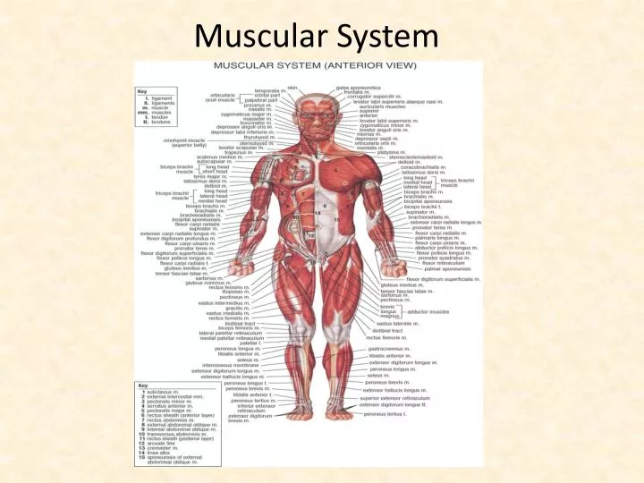
Muscular System. Lesson Objectives. 1. Compare and contrast skeletal muscles, smooth muscles, and cardiac muscles 2. Describe the structure and function of skeletal muscle 3. Compare and contrast the difference between tendons and ligaments. Objectives continued.

E N D
Lesson Objectives • 1. Compare and contrast skeletal muscles, smooth muscles, and cardiac muscles • 2. Describe the structure and function of skeletal muscle • 3. Compare and contrast the difference between tendons and ligaments
Objectives continued • 4. Describe actin and myosin’s role in the sliding filament theory • 5. Describe how acetylcholine and calcium stimulate muscle contraction • 6. Describe how troponin and tropomyosin interact to control the attachment of myosin crossbridges to actin
Skeletal Muscles • Purpose: • Location: • Structure: • Appearance:
Smooth Muscle • Purpose: • Location: • Structure: • Appearance:
Cardiac Muscle • Intercalated disk: • The fused ends of cardiac muscle cells • Purpose: • Serve as a communication device between neighboring cells • Results in sequential contraction of the cells Intercalated disk
Compare & Contrast Skeletal, Smooth, and Cardiac Muscles Skeletal Muscles -controls voluntary movements -found attached to bones -made up of fused muscle fibers -appear striated/striped -multinucleated cells Smooth Muscle -controls involuntary movements -found in walls of digestive organs, urinary bladder, arteries, veins -made up of layered sheets of cells -appear unstriated/ smooth -single nuclei Cardiac Muscle -controls involuntary contractions of the heart -found in the heart only -made up of bundles of cells -appear striated, cells branch, ends of cells fused together -single nuclei
Structure of Skeletal Muscle • Skeletal muscle is made up of fused muscle fibers • Myofibrils: are the small fibers that are bound together making up the larger muscle fibers
Skeletal Muscle Structure • The myofibrils are made up of even smaller protein filaments • 1 thick filament: myosin • 1 thin filament: actin • A myofibril consists of repeating units called sarcomeres. • Sarcomeres are where muscle contraction occurs
A closer look at the sarcomeres… • A band • Where actin & myosin overlap • I band • Contains only actin • Z line • Where adjacent actin filaments meet • Area between 2 Z lines is the sarcomere
Sliding Filament Theory-Summary • Muscle contraction causes actin to slide over myosin • As muscle contraction increases, so does the overlap between actin & myosin • This overlap causes the sarcomere to shorten (muscle contraction)
Sliding Filament Theory-Detailed • Myosin heads form crossbridges with actin filaments during muscle contraction. • When calcium & ATP are present actin binding sites are exposed
The myosin heads will move forward “walking” the actin filaments together (Power Stroke) • This walking decreases the distance between the actin & myosin. • This is the mechanism of muscle contraction
http://www.octc.kctcs.edu/gcaplan/anat/Notes/API%20Notes%20J%20%20Muscle%20Contraction.htmhttp://www.octc.kctcs.edu/gcaplan/anat/Notes/API%20Notes%20J%20%20Muscle%20Contraction.htm
Where does the calcium come from? • Calcium is stored in the sarcoplasmic reticulum • Nerves release acetylcholine to stimulate the sarcoplasmic reticulum to release calcium • Presence of calcium stimulates muscles to contract
Troponin and Tropomyosin • These 2 proteins interact to control the attachment of crossbridges to actin. • When intracellular calcium is low, tropomyosin blocks all actin sites • When intracellular calcium increases, calcium binds to troponin which in turn alters the position of tropomyosin on the actin filament. • Once tropomyosin has changed positions, the actin sites are exposed allowing myosin heads to attach to form crossbridges.
Let’s see muscle contraction at work • Tendons: Connects muscle to bone • Antagonistic Pairs • Work in opposite directions Tendon
How does this really work?? • When bending the arm • Bicep is called a reflexor because is contracting to bend a joint • Tricep is called a extensor because it is relaxed or extended • When arm is straightened • The bicep becomes the extensor • The tricep becomes the reflexor