Download
1 / 20
200 likes | 699 Views
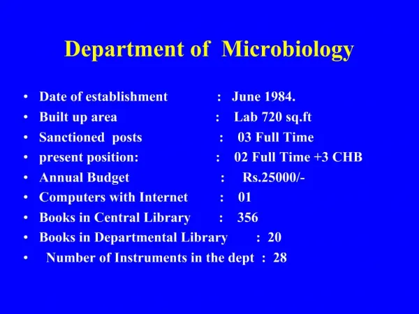
AGRICULTURAL AND ENVIRONMENTAL MICROBIOLOGY Department of Microbiology , Nutrition and Dietetics. Course syllabus – laboratory exercises Lecturer: Prof. Vojtěch Rada Office No.: 221 rada @ af.czu.cz. Course syllabus – laboratory exercises.

E N D
AGRICULTURAL AND ENVIRONMENTAL MICROBIOLOGYDepartment of Microbiology, Nutrition and Dietetics • Course syllabus – laboratory exercises • Lecturer: Prof. Vojtěch Rada • Office No.: 221 • rada@af.czu.cz
Course syllabus – laboratory exercises • Microscopyofbacteria: Negative staining, simplestaining • Microscopyofbacteria: Gram staining • Yeast study: Methylenebluestaining • Mould study: Zygomycetes (Mucor, Rhizopus), Ascomycetes (Eupenicillium) and Deuteromycetes (Penicillium, Aspergillus. Asexual reproduction (conidiospores and sporangiospores) • Actinomycetesandantibiotics
Course syllabus – laboratory exercises • Identification of bacteria (staphylococci) • Microbiology of drinking water • Microbiology of milk: fermented milk products, starter cultures. • Carbon cycle: amylolytic bacteria • Nitrogen cycle: Nitrogen fixing bacteria (Azotobacter, Rhizobium)
LABORATORY SAFETY • Do not drink, eat and smoke • Protective clothing • Aseptic technique • Bacteriological loop, needle • Bunsen burner • Bacteriological stains
Brightfield microscopy • low-power objectives (4-20x) • high-dry objectives (40-60x) • immersion objectives (90-100x) • Resolution limit (0.4 μm) • Magnification(1500x) • Oil immersion technique.
PROTOZA 100 μm MOULDS AND YEASTS 5 – 10 μm BACTERIA - COCCI 1 μm - RODS 1 x 2 – 4 μm VIRUSES 0,1 μm
Methylene blue staining • This method distinguish live (colourless) and dead (coloured) cell. • Saccharomyces cerevisiae – yeast; baker, beer production
Methylene blue staining • A drop of water is placed in the centre of a slide. • Two loopful of yeast are transferred to slide • One loopful of methylene blue is added • Examine with dry objectives
YEASTS budding
COCCI pediococci, tetrades diplococci sarcina streptococci staphylococci
Axis of division diplococci streptokoky tetrades sarcinas staphylococci
RODS- regular endospore-forming plektridium Clostridium Bacillus
RODS - curved vibrio spirilla spirochaeta
SPIROCHAETA VIBRIO
RODS- irregular bifidobacteria mycobacteria
Negative Staining • (Background staining) • This method consist of mixing the microorganisms in a small amount of nigrosine and spreading the mixture over the surface of a slide.
Negative Staining • Drops of water and nigrosine are placed in the centre of a microscopic slide. • Remove a small amount of material from between your teeth with a sterile straight toohpick. • Spread the mixture of water, nigrosine and sample over the slide. • Allow the slide to air-dry and examine with an oil immersion objective