Download
1 / 65
650 likes | 791 Views
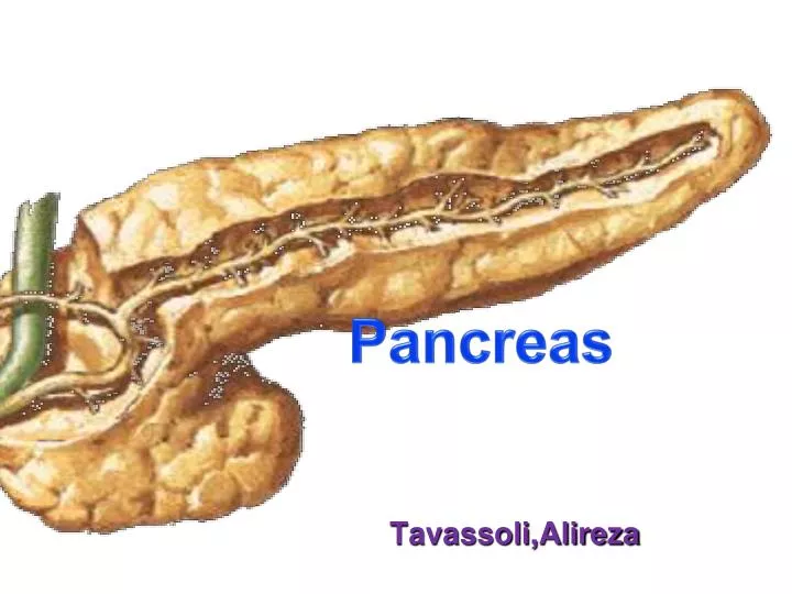
Pancreas . Tavassoli,Alireza. نام درس :بيمار يهاي پانكراس اهداف. 1-بيماري هاي پانكراس رابشناسد 2-عوامل سبب زاي بيماري هاي پانكراس را بشناسد 3- راه هاي پيشگيري بيماري هاي پانكراس را بداند . 4- نحوه مراقبت بيماري هاي پانكراس را فراگيرد . (Surveillance)

E N D
Pancreas Tavassoli,Alireza
نام درس :بيمار يهاي پانكراساهداف • 1-بيماري هاي پانكراس رابشناسد • 2-عوامل سبب زاي بيماري هاي پانكراس را بشناسد • 3- راه هاي پيشگيري بيماري هاي پانكراس را بداند. • 4- نحوه مراقبت بيماري هاي پانكراس را فراگيرد. (Surveillance) • 5-عوامل خطرزاي بيماري هايپانكراس را بداند • 6- ويژگي هاي اپيدميولوژيك بيماري هاي پانكراس را بشناسد . • 7-روش هاي تشخیصی بيماري هاي پانكراس رابشناسد. • 8- روش هاي درمان را بداند.
neutralize chyme digestive enzymes hormones Normal Anatomy & Physiology
Exocrine Function BODY common bile duct TAIL HEAD pancreatic duct ampulla UNCINATE pancreatic enzymes
Enzyme Secretion acinus pancreatic duct microscopic view of pancreatic acini duodenum
Exocrine Stimulation • The moreproximal the nutrient infusion…the greater the pancreatic stimulation (dog studies) • stomach – maximal stimulation • duodenum – intermediate stimulation • jejunum – minimal / negligible stimulation • Elemental formulas tend to cause less stimulation than standard intact formulas • intact protein > oligopeptides > free amino acids • Intravenous nutrients (even lipids) do not appear to stimulate the pancreas
Clinical Case • A man with acute onset abdominal pain • h/o alcohol intake • Or Gall stone
Acute PancreatitisDefinition • Acute inflammatory process involving the pancreas • Usually painful and self-limited • Isolated event or a recurring illness • Pancreatic function and morphology return to normal after (or between) attacks
Cholelithiasis Ethanol abuse Idiopathic Medications Hyperlipidemia ERCP Trauma Pancreas divisum Hereditary Hypercalcemia Viral infections Mumps Coxsackie virus End-stage renal failure Penetrating peptic ulcer Acute PancreatitisAssociated Conditions
Acute PancreatitisCausative Drugs • AIDS therapy:pentamidine ,didanosine • Anti-inflammatory:sulindac, salicylates • Antimicrobials:metronidazole, sulfonamides, tetracycline, nitrofurantoin • Diuretics:furosemide, thiazides • IBD:sulfasalazine, mesalamine • Immunosuppressives:azathioprine, 6-mercaptopurine • Neuropsychiatric:valproic acid • Other: calcium, estrogen, tamoxifen,
Hereditary Pancreatitis • Autosomal dominant with 80% phenotypic penetrance • Recurrent acute pancreatitis, chronic pancreatitis, and 50-fold increased risk of pancreatic cancer
PancreatitisBackground • Potentially fatal • Mortality – 10-15% • Necrosis determines the prognosis
Background • Mild AP (no necrosis) – 0% • Sterile necrosis – 10% • Infected necrosis – 25% • Overall mortality: 10-15%
What do you think? • Amylase or lipase • Ultrasound or CT scan • If yes, When? • ICU or medical ward • Enteral nutrition or TPN • Antibiotics • ERCP • Surgery
prematureenzyme activation Acute PancreatitisPathogenesis acinar cell injury failed protectivemechanisms
Acute PancreatitisPathogenesis premature enzyme activation autodigestion of pancreatic tissue local vascular insufficiency activation of white blood cells release ofenzymes into the circulation localcomplications distantorgan failure
Acute PancreatitisPathogenesis SEVERITY Mild Severe • STAGE 1: Pancreatic Injury • Edema • Inflammation • STAGE 2: Local Effects • Retroperitoneal edema • Ileus • STAGE 3: Systemic Complications • Hypotension/shock • Metabolic disturbances • Sepsis/organ failure
PancreatitisClinical Presentation • Pain:Steady & severe in nature; located in the epigastric or umbilical region; may radiate to the back. Worsened by lying supine; may be lessened by flexed knee, curved-back position. • Vomiting:Varies in severity, but is usually protracted, worsened by ingestion of food or fluid. Does not relieve the pain. Usually accompanied by nausea.
Pancreatitiscon’t…… • Fever: Rarely exceeds 39 C. • Abdominal Finding:Rigidity, tenderness, guarding, distended Abd, decreased or absent peristalsis and paralytic ileus.Fatty stools-(steatorrhea) • Laboratory Finding:Elevation of WBC count- 20-50,000. lipase and amylase(5 to 40 times); elevated(glucose, bilirubin, alkaline phosphatase.,Urine amylase).Abnormal low serum CA, Na & Mg.-due to dehydration. Binding of Ca in areas of fat necrosis.
Acute Pancreatitis
Acute Pancreatitis
Acute Pancreatitis
Acute Pancreatitis
Acute PancreatitisDifferential Diagnosis • Choledocholithiasis • Perforated ulcer • Mesenteric ischemia • Intestinal obstruction • Ectopic pregnancy
Acute PancreatitisDiagnosis • Symptoms & Signs • Abdominal pain • Laboratory • Elevated amylase or lipase • > 3x upper limits of normal • Imaging • Abnormal sonogram or CT
Acute PancreatitisDiagnosis • EtOH: history • Gallstones: abnormal LFTs & sonographY • Hyperlipidemia: lipemic serum, Tri>1,000 • Hypercalcemia: elevated Ca • Trauma: history • Medications: history
Abdominal Exam • Abdominal tenderness and rigidity • Bowel sounds decreased • Palpable upper abdominal mass Acute fluid collections and pseudocysts • Skin Exam • Erythematous skin Nodule (Subcutaneous Fat Necrosis) • Cullen's Sign (periumbilical discoloration) • Turner's Sign (flank discoloration) * due to exudation of blood-stained fluid into the subcutaneous tissue, usually 72 h into the illness.
Acute PancreatitisClinical Manifestations PANCREATIC PERIPANCREATIC Adjacent viscera: SYSTEMIC Mild: edema, inflammation, fat necrosis Severe:phlegmon, necrosis, hemorrhage, infection, abscess, fluid collections Retroperitoneum, perirenal spaces, mesocolon, omentum, and mediastinum ileus, obstruction, perforation Cardiovascular: hypotension Pulmonary: pleural effusions, ARDS Renal: acute tubular necrosis Hematologic: disseminated intravascular coag. Metabolic:hypocalcemia, hyperglycemia
Serum Amylase elevated Nonspecific Returns to normal in 48-72 hours Normal amylase does not exclude pancreatitis Level of elevation does not predict disease severity Serum Lipase elevated Specific for pancreatic disease Returns to normal in 7-14 days Diagnosis: Biochemical
White Blood Cells increased to 15k-20k Hypertriglyceridemia (15%) liver Function Tests (ALP) (AST) ,elevated (LDH) elevated (Poor prognosis) Hyperglycemia Albumine (Poor prognosis) Serum Electrolytes Hypocalcemia (25%) Diagnosis: Biochemical
Another criteria often used to assess the severity of pancreatitis is the (APACHE-II) . Acute Physiology And Chronic Health Evaluation age and vital signs Specific laboratory parameters, Chronic health status The main advantage is the immediate assessment of the severity of pancreatitis. A score of eight or more at admission is usually considered indicative of severe disease
Predictors of Severity • Why are they needed? • Appropriate triage & therapy • compare results of studies of the impact of therapy • When are they needed? • optimally, within the first 24 hours • Which is the best?
AT ADMISSION Age > 55 years WBC > 16,000 Glucose > 200 AST > 250 IU/L LDH > 350 IU/L WITHIN 48 HOURS HCT drop > 10% BUN > 5 Arterial PO2 < 60 mm Hg Base deficit > 4 mEq/L Serum Ca < 8 Fluid sequestration > 6L Ranson CriteriaAlcoholic Pancreatitis Number <2 1% 3-4 16% 5-6 40% 7-8 100% Mortality
Glasgow CriteriaNon-alcoholic Pancreatitis • WBC > 15,000 • Glucose > 180 • BUN > 16 • Arterial PO2 < 60 mm Hg • Ca < 8 • Albumin < 3.2 • LDH > 600 U/L • AST or ALT > 200 U/L
CT Severity Index Balthazar et al. Radiology 1990.
Useful markers of severe disease. • Pleural effusion • BMI (High body mass index) • Necrosison contrast-enhanced CT-SCAN • CRPlevel greater than 150 mg/L at 48 h • Infection of the necrotic tissue after the first week of illness is the major determinant of later outcome.
CT-guided percutaneous fine-needle aspiration of the pancreatic tail