Download
1 / 27
420 likes | 915 Views
Carbohydrate Metabolism. Digestion & Absorption of Carbohydrates. 1- In the Mouth Salivary amylase digests starch partially into mixtures of dextrins and maltose. 2- In the Stomach

E N D
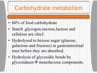
Digestion & Absorption of Carbohydrates 1- In the Mouth Salivary amylase digests starch partially into mixtures of dextrins and maltose. 2- In the Stomach Salivary amylase acts for short time till the gastric HCL inhibits the enzyme (due to drop of pH). Small amount of acid hydrolysis occurs in stomach. 3- In the Small Intestine a- Pancreatic amylase It completes the digestion of starch to maltose and isomaltose. b- Intestinal disaccharidases They complete the action of other enzymes with production of monosaccharides e.g. sucrase, maltase, lactase acting on sucrose, maltose,lactose.
So, the end products of carbohydrate digestion are mainly glucose, galactose and fructose. • Monosaccharides are then absorbed actively by small intestine. • The portal vein carries simple sugars to the liver where they are metabolized. • Liver does not consume all sugars passing through it, but a large proportion of these sugars is deliverded to systemic blood in order to be utilized by other tissues.
Summary Diagram for Fate of Absorbed Sugars Fructose Glucose Glycerol 3-phosphate Galactose FA Oxidation Glycogen Glycerophosphatides Carbon skeleton of Amino acids Other carbohydrates TAG Cholesterol & other steroids
Oxidation of glucose A- The Major Pathways for Oxidationwhich are mainly concerned with energy production. I- Glycolysis: It produces pyruvate under aerobic condition and lactate under anaerobic condition. II- Citric acid cycle (Krebs’ cycle):Under aerobic condition, pyruvate is converted to active acetate for oxidation through citric acid cycle. B- The Minor Pathways for Oxidationwhich are mainly for synthesis of other glucose derivatives and not for energy production. I- Hexosemonophosphate pathway (HMP): For production of pentoses and NADPH. II- Uronic acid pathway: For production of uronic acids.
A- The Major Pathways for Oxidation I-GLYCOLYSIS • Sequence of enzymatic reactions in which one molecule of glucose is converted into two molecules of three carbon compound, either pyruvatein the presence of oxygen or lactate in the absence of oxygen. • Glycolyticpathway proceeds in the cytosol of all cells (all of the enzymes of glycolysis are found in the cytosol). • Glycolysisis also termed anaerobic oxidation of glucose as it can proceed in the absence of oxygen.
Steps of Glycolysis can be divided into two phases: Phase I In this preparatory stage, glucose is phosphorylated and cleaved to yield two molecules of glyceraldehyde 3-phosphate. This process consumes 2 ATP. Phase II The two molecules of glyceraldehyde 3-phosphate are converted to pyruvate under aerobic state with generation of 4 ATP at substrate level and 6 ATP at the respiratory chain level. Under anaerobic state only 4 ATP are formed at the substrate level with conversion of pyruvate to lactate.
Regulation of Glycolysis: Glycolysis is regulated at three nonequilibrium (irreversible) reactions, i.e. 3 key enzymes: • Glucokinase(or hexokinase), • Phosphofructokinase-1(PFK-1) • Pyruvatekinase(PK).
II- CITRIC ACID CYCLE (Tricarboxylic Acid Cycle or Krebs' Cycle) • It is formed of a series of reactions that are responsible for the complete oxidation of the acetyl moiety of acetyl-CoA. • It is the final common pathway for the oxidation of carbohydrates, lipids and proteins because glucose, fatty acids and most amino acids are metabolized to acetyl-CoA or intermediates of the cycle. • During the oxidation of acetyl-CoA, coenzymes (NAD and FAD) are reduced and subsequently reoxidized in the respiratory chain with the formation of ATP.
Site: • The enzymes of the TCA cycle are found in the mitochondrial matrix except succinatedehydrogenasewhich is tightly bound to the inner mitochondrial membrane (forms complex II of the respiratory chain). • The enzymes of the TCA cycle are in close proximity to the enzymes of the respiratory chain.
B- The Minor Pathways for Oxidation I- HexoseMonophosphate Pathway (HMP): The pentose phosphate pathway (PPP) is an alternative route for glucose oxidation. It has two major functions, formation of: 1) Ribose-5-p required for nucleotide and nucleic acid synthesis 2) NADPH The enzymes of the pentose phosphate pathway are cytosolic. HMP is active in certain tissues e.g. liver, thyroid, adrenal cortex, adipose tissue, gonads, retina, lactating mammary gland and RBCs.
NADPH is required for the following reactions: • Reduction of metabolically impotant compounds as glutathione and folic acid. • Synthesis of fatty acids • Cholesterol synthesis • Hydroxylation reactions. • Glutathione is of particular importance to combat oxidation stress in tissues and to keep hemoglobin active by conserving iron in ferrous state
II-Uronic Acid Pathway Uronic acid pathway is also an alternative oxidative pathway for glucose that does not lead to the formation of ATP. Uronic acid pathway is a cytosolic pathway that occurs in the liver. In humans, it catalyzes the conversion of glucose to glucuronic acid, and pentoses. Importance of Uronic acid pathway The main function is the formation of UDP-glucuronate which is utilized in the following pathways: • Glycosaminoglycans(GAGs) synthesis. • Conjugation reactions with many compounds to increase their water solubility such as: all steroid hormones and their metabolites, bilirubin and certain detoxification reactions of xenbiotics such as phenols.
Glycogen Metabolism • Glycogen is a highly branched polymer of glucose. It is the main storage form of carbohydrates in animals. It is present mainly in the liver and in muscles. • Liver glycogen (forms 8 – 10% of its wet weight) maintains blood glucose between meals. After 12–18 hours of fasting, liver glycogen is almost totally depleted. • Muscle glycogen(forms 2% of its wet weight) :Muscle glycogen supplies the contracting muscles with a readily available source of glucose.
Glycogen metabolism includes the following: I- Glycogenesis Glycogenesis is synthesis of glycogen from glucose-6-p.this requires presence of the enzyme glycogen synthase. II- Glycogenolysis Glycogenolysis is breking-down of glycogen to glucose (in the liver) or to g-6-p (in the muscle) Both glycogenesis and glycogenolysis are under strict hormonal control mediated by a second messenger cyclic AMP
GLUCONEOGENESIS • It is the synthesis of glucose and /or glycogen from non-carbohydrate precursors e.g. glycerol, glucogenic amino acids and lactate. • Its main function is to supply blood glucose in case of carbohydrate deficiency (fasting, starvation and low carbohydrate diet). • It starts 4 to 6 hours after the last meal and continues throughout fasting state.
Regulation of Blood Glucose • The normal fasting plasma glucose level (after 8-12 hours fasting) is between 70 to less than 100 mg/dL, increases after meal and returns back to <140 mg/dL at two hours after feeding (2 hour postprandial or 2h PP). • The maintenance of blood glucose is an important function of different tissues. Glucose is the principal source for energy production in the brain. • Sudden decrease in blood glucose if not treated may produce coma or even death.
After-meal rise in blood glucose stimulates insulin secretion from pancreatic β-cells of islets of langerhans. Insulin action: It is secreted by the B-cells of pancreatic islets in response to hyperglycemia. It produces its effects through the following mechanisms: • It increases the uptake of glucose by extrahepatic tissues (heart, skeletal muscles and adipose tissues). • It increases utilization of glucose (oxidation, glycogenesis and lipogenesis) in different tissues. • It decreases output of glucose by liver (decreases glycogenolysis and gluconeogenesis). During fasting blood glucose decreases so insulin secretion is inhibited whereas the anti-insulin hormones increase leading to activation of mechanisms of glucose production: glycogenesis and gluconeogenesis.
During fasting blood glucose decreases so insulin secretion is inhibited whereas the anti-insulin hormones increase leading to activation of mechanisms of glucose production: glycogenesisandgluconeogenesis.
Diabetes Mellitus Definition The term diabetes mellitus describes a metabolic disorder that is characterized by persistent rise in blood glucose (hyperglycemia) result from defects in insulin secretion, insulin action, or both. Classification of DM 1) Type I Diabetes Mellitus Type I diabetes is primarily a disease of the young. It was previously known as insulin dependent diabetes mellitus (IDDM) means that it necessitates insulin to control hyperglycaemia 2) Type II Diabetes Mellitus It is previously known as noninsulin dependent diabetes mellitus (NIDDM) means that drugs stimulate endogenous insulin secretion and promoting glucose utilization are required.
Metabolic Changes in DM All the metabolic changes are due to decrease in the insulin / anti-insulin ratio, which produces changes reversal to insulin action or as a consequent of hyperglycemia. 1) Changes in carbohydrate metabolism include: This leads to hyperglycemia, glucosuria, polyuria, loss of electrolytes, dehydration, and polydepsia. 2) Changes in lipid metabolism include: Decreased lipogenesis and increased lipolysis. This leads to weight loss and increases plasma free fatty acids. 3) Changes in protein metabolism include: it leads to increased sensitivity to infection and delayed healing of wounds.
Complications of DM: • These complications can occur over a long period of time. • They can be divided in macrovascular and microvascular complications. • Diabetes accelerates atherosclerosis that can lead to coronary artery disease, stroke and peripheral vascular disease (macrovascular disease) • Damage to the retina (retinopathy), kidney (nephropathy) and nerves (neuropathy) (microvascular disease).
Types of Diabetic Coma I- Diabetic Ketoacidosis: • Diabetic ketoacidosis is considered a medical emergency that results from uncontrolled hyperglycemia and deficiency of insulin. • The condition can be precipitated by stress and infection. Diabetic ketoacidosis is much more common in type I diabetes, but can also occur in patients with type II diabetes. II- Hypoglycemic Coma • Hypoglycemia results from taking too much diabetes medication or insulin. • It is manifested as headache, feeling dizzy, poor concentration, tremors of hands, and sweating are common symptoms of hypoglycemia. Coma occurs if blood sugar level gets too low.
Diagnosis of DM: • Many patients with diabetes remain asymptomatic for long periods, so that the first presentation of the disease is frequently a chronic complication. • Symptoms include polyuria, polydipsia, polyphagia, prolonged time of wound healing, and weight loss. • Polyphagiaresults from the decreased glucose uptake by the satiety centre in the brain.
Tests for Diagnosis and Assessment of DM Control I- Fasting and 2-hour (post-glucose or postprandial) plasma glucose levels in an oral glucose tolerance test (OGTT). Non-diabetic healthy subjects will have: Fasting plasma glucose < 100 mg/dL, Two-hours value in an OGTT (2-h PG) < 140 mg/dL. Patients with diabetes mellitus will have: Fasting plasma glucose > 126 mg/dL, 2-hour value in an OGTT (2-h PG) at or above 200 mg/dL. II- Oral Glucose Tolerance Test (OGTT) Normal OGTT: There is an increase in plasma glucose levels after 30 and 60 minutes from the glucose load due to the absorption of glucose followed by a drop due to increased uptake and utilization of glucose under the effect of the stimulated insulin secretion. No glucosuria occurs during the test.
III- Measurement of Glycated- Hb (HbA1C): It is a good for diagnosis and monitor of blood glucose to assess diabetic control and to follow up of diabetic patients. Normal HBA1C is 4 – 6.5%; levels above 6.5% are diagnostic of diabetes mellitus and levels > 8 % indicate poor diabetic control.
Treatment of Diabetes Mellitus • Diet control It is to achieve weight reduction in overweight patients with type II DM. If improvement in hyperglycemia is not achieved by diet, trial with an oral drug should be started. • Oral Anti-diabetic Drugs: These drugs are used for type II DM but not for type I DM oral hypoglycemic drugs are: Drugs that increase insulin secretion , improve insulin sensitivity or decrease the intestinal absorption of carbohydrates and fats • Insulin: Human insulin is now available in the market and often it is preferred. It is injected subcutaneously, or as an insulin pen. Recently, inhaled insulin is under trials.