Download
1 / 25
250 likes | 434 Views
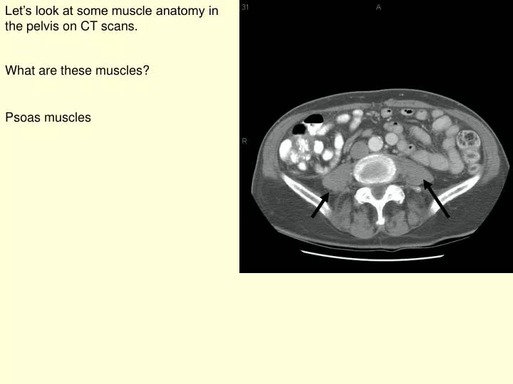
Let’s look at some muscle anatomy in the pelvis on CT scans. What are these muscles?. Psoas muscles. Which are the muscles that you are starting to see in front of and behind the iliac bones?. Gluteus medius. Iliacus. Can you idendify the abdominal muscles at this level?. Rectus abdominis.

E N D
Let’s look at some muscle anatomy in the pelvis on CT scans. What are these muscles? Psoas muscles
Which are the muscles that you are starting to see in front of and behind the iliac bones? Gluteus medius Iliacus
Can you idendify the abdominal muscles at this level? Rectus abdominis External oblique (superficial) Internal oblique (middle) Transversus abdominis (deep)
What muscle are we starting to get into just posterior to the gluteus medius? This is the gluteus maximus (it doesn’t look very maximus at this level it gets larger more inferiorly)
At this level you start to see the iliacus and the psoas coming together and we also start to get into the third gluteal muscle. This is the gluteus minimus
Which muscles are these? Piriformis muscles Which major nerve is this? Sciatic nerve This is where the sciatic nerve comes out of the greater sciatic foramen. What are the anatomic boundaries of the greater sciatic foramen and what comes out of it?
What is the name of this muscle? Sartorius What is the origin of this muscle? Anterior superior iliac spine. You can see it if you follow the muscle 4 slices superiorly.
Which muscle is this? Hint, it is the only member of the quad group to cross both the hip and the knee joint. Rectus femoris What is the origin of the rectus femoris? Anterior inferior iliac spine, which you can see if you trace the muscle 2 images superiorly.
What portion of the bony pelvis is the arrow pointing to? Hint you are sitting on it right now. Ischial tuberosity Which flexor of the knee attaches here? The hamstrings
Which is this lateral muscle? Hint, if you trace it back it has the same attachment point as the sartorius on the anterior superior iliac spine. Tensor fascia lata
This is a normal x-ray of the hips. Not the joint space between the femoral heads and the acetabulae.
This is a patient with osteoarthritis in the left hip. Not there is no joint space in the superior aspect of the hip joint. There is also sclerosis (increased density in the bone) above the joint space characteristic of osteoarthritis. There is also osteophyte formation (bony production on the edges of the joint-arrow). This is also commonly seen in osteoarthritis.
These are 2 different types of hip prostheses in this patient with bilateral total hip replacements.
This is a normal view of the pelvis. When looking for acetabular fractures there a few lines to look at. This is the iliopubic line which outlines the anatomic anterior column This is the ilioischial line which outlines the anatomic posterior column.
This is a Judet view (oblique view) of the pelvis which better illustrates the columns of the acetabulum. Can you find the anterior column (iliopubic line)? Can you identify the posterior wall
This is the opposite oblique. Can you find the region of the posterior column? Can you identify the anterior wall/lip of the right acetabulum
This is a case with an acetabular fracture. Do you see disruption of one of the columns? The anterior column is disrupted
This is another oblique view. Do you see disruption of one of the columns? The posterior column is disrupted
Anterior Posterior On the left you see a view looking from the outside of the pelvis. The acetabulum is the dark structure in the middle (arrow). The fracture line shows how a posterior column fracture runs On the right you see a sagittally reformatted CT (oriented the same way as the anatomic drawing to show the fracture line of a posterior column fracture.
Anterior division Posterior division These are consecutive sagittal images illustrating the anterior and posterior divisions of the internal iliac artery. Can you try and identify the superior and inferior gluteal arteries.
This is a 3D reformatted images of the CT angiogram from the previous slide. The arrow points to the posterior division of the internal iliac artery. Can you see why the superior gluteal artery could be injured with a posterior column fracture? Fracture plane of a posterior column fracture