Download
1 / 64
650 likes | 1.27k Views
Atrial & Junctional Dysrhythmias. Terry White, RN, EMT-P. Atrial Premature Atrial Complex Wandering Atrial Pacemaker Atrial Tachycardia (ectopic) Multifocal Atrial Tachycardia Atrial Flutter Atrial Fibrillation. Junctional Junctional Escape Rhythm Premature Junctional Complex

E N D
Atrial & Junctional Dysrhythmias Terry White, RN, EMT-P
Atrial Premature Atrial Complex Wandering Atrial Pacemaker Atrial Tachycardia (ectopic) Multifocal Atrial Tachycardia Atrial Flutter Atrial Fibrillation Junctional Junctional Escape Rhythm Premature Junctional Complex Junctional Tachycardia Accelerated Junctional Rhythm AV Nodal Re-entrant Tachycardia (PSVT) Atrial & Junctional Dysrhythmias
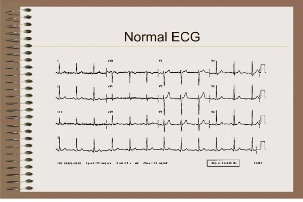
Atrial & Junctional vs. SA Node • Origin of the pacemaker site is at or above the AV junction but is not the SA Node • Single Atrial site • Multiple atrial sites • AV Junction • Common Characteristics • Narrow QRS • Without regular, typical appearing, discernible P waves • Regular or Irregular Rhythm
Premature Atrial Complex (PAC) • PAC - Ectopic beat from the Atria • earlier than expected • Complex, Not a rhythm! • Assess the underlying rhythm first
Premature Atrial Complex (PAC) • Causes • Idiopathic • Caffeine, tobacco, alcohol • Stress, Emotion, Infection • Digitalis toxicity • Hypoxia • Congestive failure • Increased sympathetic tone
Premature Atrial Complex (PAC) • Characteristics • Heart Rate: dependent on the underlying rhythm • Rhythm: irregular if PACs are present; underlying rhythm may be regular • PacemakerSite: ectopic site in the atria; underlying rhythm has its own pacemaker site • P Waves: earlier than next expected P wave; positive in lead II; may not look like other P waves present • P-R Interval: usually normal for the PAC • R-R Interval: unequal since PACs present • QRS Complex: usually narrow • P to QRS: usually one to one relationship
Premature Atrial Complex (PAC) • Characteristics • Paired Ectopic Beats referred to as couplet • Alternating Ectopic Beat referred to as Bigeminy, Trigeminy, or Quadrigeminy • e.g. Atrial Bigeminy or Ventricular Bigeminy • May not always result in ventricular conduction • “Blocked PAC” or “Non-conducted PAC” • No compensatory pause in PAC • Compensatory vs. Noncompensatory Pause
Compensatory vs Noncompensatory Pause • Compare the distance between 3 normal beats • Noncompensatory • the normal beat following the premature complex occurs before it was expected (the distance not the same) • Compensatory • the normal beat following the premature complex occurs when expected (the distance is the same)
Premature Atrial Complex (PAC) • Management • Usually not clinically significant • treat underlying cause • Frequent PACs may indicated enhanced automaticity of atria or reentry mechanism • may warn of or initiate supraventricular arrhythmias such as atrial tachycardia, atrial flutter, atrial fibrillation or PSVT • if nonconducted PACs are frequent and HR < 50, treat as bradycardia • PACs may be wide (aberrant conduction) and must be differentiated form PVCs
Wandering Atrial Pacemaker • Pathophysiology • shifting of pacemaker focus from one to another within the atrial tissue • May be associated with ischemic disease involving the sinus node or an inflammatory state (e.g. rheumatic fever) • May occur without any finding of disease
Wandering Atrial Pacemaker • Characteristics • Heart Rate: usually 60-100 bpm • Rhythm: irregularly irregular (one of three) • PacemakerSite: variable, all within the atria including SA node • P Waves: variable including normal appearing P waves • P-R Interval: unequal, varies • R-R Interval: unequal, varies • QRS Complex: usually narrow • P to QRS: usually one to one relationship
Wandering Atrial Pacemaker • Management • ECG rhythm generally does not require treatment • Underlying cause may require treatment
Multifocal Atrial Tachycardia • Pathophysiology • Same as WAP just faster than 100 bpm • An uncommon ECG rhythm • Usually seen in someone with COPD or severe systemic disease (e.g. sepsis, shock)
Multifocal Atrial Tachycardia • Characteristics • Heart Rate: >100 bpm • Rhythm: irregularly irregular (one of three) • PacemakerSite: variable, all within the atria including SA node • P Waves: variable including normal appearing P waves • P-R Interval: unequal, varies • R-R Interval: unequal, varies • QRS Complex: usually narrow • P to QRS: one to one relationship
Multifocal Atrial Tachycardia • Management • Treated like Supraventricular Tachycardia
Tachycardia Management Overview • If Unstable : • Immediate Synchronized Cardioversion! • If Stable: • IV/O2/Monitor/12 lead • Identify Rhythm using 12 lead if necessary • Drug therapy • If drugs fail, then synchronized cardioversion
Tachycardia: Narrow Complex • Primary/Secondary ABCD • Vagal maneuvers • Adenosine 6 mg rapid IV push, with flush • Repeat with 12 mg rapid IV push with flush • Other Considerations • amiodarone 150 mg slow IV (15 mg/min) • procainamide 20-30 mg/min IV • diltiazem 0.25 mg/kg slow IV or verapamil 2.5 mg slow IV if NO WPW/Hypotension • synchronized cardioversion
Atrial Flutter • Signature • “Saw tooth” baseline • Commonly occurs in multiples • 300, 150, 75 • based on degree of AV block
Atrial Flutter • Causes • Myocardial ischemia • Hypoxia • CHF • COPD (cor pulmonale) • Hyperthyroidism • Digitalis toxicity • Not a common dysrhythmia
Atrial Flutter • Characteristics • Heart Rate: usually multiples - 300, 150, 75 • Rhythm: usually regular except with variable AV block • PacemakerSite: atrial site • P Waves: No P waves; Flutter (F) waves • P-R Interval: not applicable • R-R Interval: usually equal except with variable AV block • QRS Complex: usually narrow • P to QRS: not applicable
Atrial Fibrillation (A-Fib) • Signature • Irregularly irregular • No organized atrial activity • Types • A-Fib with uncontrolled ventricular response (rate > 100, usually 160-180) • A-Fib with controlled ventricular response(rate < 100, usually 60-70)
Atrial Fibrillation • Characteristics • Heart Rate: atrial rate may be very fast, avg of 400 bpm; variable ventricular rate • Rhythm: irregularly irregular • PacemakerSite: multiple atrial sites • P Waves: No P waves; fibrillation (f) waves • P-R Interval: not applicable • R-R Interval: usually unequal • QRS Complex: usually narrow • P to QRS: not applicable
Atrial Fibrillation • Causes • Myocardial ischemia • Hypoxia • CHF • COPD (cor pulmonale) • Hyperthyroidism • Digitalis toxicity • Idiopathic
Atrial Fibrillation • Presentation • Paroxysmal • Acute • Chronic
Atrial Fibrillation • Complications • Loss of atrial kick • Thrombus formation • Emboli
Tachycardia: A.fib/A. flutter • Primary/Secondary ABCD • Assess for WPW • No WPW • Calcium channel blockers • WPW • amiodarone 150 mg slow IV (15 mg/min) • procainamide 20 –30 mg/min IV
Atrial Fib/Flutter Treatment • Rapid Response/Stable with Symptoms • Oxygen, Monitor, IV • Vagal maneuvers (if needed as a diagnostic tool) • No WPW • Verapamil, 2.5 - 5 mg slow IV over 2 min, may repeat in 15-30 mins • OR, Diltiazem, 0.25 mg/kg slow IV over 2 min, may repeat i15 min at 0.35 mg/kg slow IV • Calcium channel blockers • WPW • amiodarone 150 mg slow IV (15 mg/min) • procainamide 20 –30 mg/min IV
Atrial Fib/Flutter Treatment • Rapid Response/Unstable • Oxygen, Monitor, IV • Sedate • Cardioversion • Consider anticoagulation first
Atrial Fib/Flutter Treatment • Slow Response/Unstable (usually occurs in A-Flutter) • Oxygen, Monitor, IV • Atropine • Pacemaker • Dopamine or epinephrine infusion
Atrial Fib/Flutter Treatment • Normal (controlled) Rate • Oxygen, Monitor, IV • Evaluate, treat underlying problems • Patient may have CHF with pulmonary edema or Acute MI
Supraventricular Tachycardia (SVT) • Supraventricular origin that is: • Not a sinus rhythm • Not atrial fibrillation or flutter • Not WAP or MAT • often segregated into • Nonparoxysmal Atrial Tachycardia (ectopic) • Paroxysmal Supraventricular Tachycardia (reentry) • Very often can not distinguish between the two
Supraventricular Tachycardia • Nonparoxysmal Atrial Tach • Enhanced automaticity • Patient cannot pinpoint onset • Often caused by digitalis toxicity
Supraventricular Tachycardia • Characteristics of Nonparoxysmal Atrial Tach • Heart Rate: usually 160-240 • Rhythm: regular • PacemakerSite: one ectopic atrial site • P Waves: present but not appearing as normal P waves, similar to each other, may not be easily identifiable • P-R Interval: not applicable • R-R Interval: usually equal • QRS Complex: usually narrow • P to QRS: if P waves visible, one to one relationship
Supraventricular Tachycardia • Nonparoxysmal Atrial Tach • Management • Correct underlying cause if possible • If hemodynamically unstable: • consider immediate cardioversion • If hemodynamically stable, consider: • Diltiazem, 0.25 mg/kg slow IV over 2 min, may repeat in 15 mins at 0.35 mg/kg slow IV • Metoprolol, 5 mg slow IV over 2-5 mins, may repeat in 5 min • Amiodarone, 150 mg IV infusion over 10 mins
Supraventricular Tachycardia • Paroxysmal Supraventricular Tachycardia (PSVT) • Causes • reentry mechanism at AV junction with or without an accessory pathway • onset may occur due to • increased sympathetic tone • stimulant use • electrolyte abnormalities • anxiety/emotional stress • Clinical significance dependent on rate and underlying cardiac function
Supraventricular Tachycardia • Paroxysmal Supraventricular Tachycardia (PSVT) • Episodes begin/end suddenly • Healthy patients c/o palpitations • Patients with heart disease c/o • Weakness • Dizziness • Shortness of breath • Chest pain • Pulmonary edema
Supraventricular Tachycardia • Characteristics of Paroxysmal SVT • Heart Rate: usually 160-240 • Rhythm: regular • PacemakerSite: one ectopic atrial site • P Waves: usually not identifiable • P-R Interval: not applicable • R-R Interval: usually equal • QRS Complex: usually narrow • P to QRS: not applicable
Supraventricular Tachycardia • Management • Oxygen, Monitor, IV • Assess for Stable vs Unstable • If Unstable • Immediately cardiovert
Supraventricular Tachycardia • Assess for Stable vs Unstable (cont) • If Stable • Vagal maneuvers • Avoid in digitalis toxicity • May produce AV blocks or asystole • Adenosine • 6 mg RAPID IV push, may repeat in 1-2 minutes at 12 mg RAPID IV push, then 12 mg RAPID IV push • follow each dose immediately with a 10-20 cc flush • Blocks conduction through AV node • May produce transient aystole • Short half-life (<6 seconds) • Drug Interactions
Supraventricular Tachycardia • Assess for Stable vs Unstable (cont) • If Stable PSVT remains after Adenosine and vagal maneuver, may consider: • Beta blocker • Metoprolol, 5 mg slow IV over 2-5 mins, may repeat in 5 min • ONLY if NO history of heart disease or CHF • Diltiazem • 0.25 mg/kg slow IV over 2 min, may repeat in 15 mins at 0.35 mg/kg slow IV • Amiodarone • 150 mg IV infusion over 10 mins
Synchronized Cardioversion • Sedate, if possible • Valium 5 to 10 mg IV, or • Versed 2.5 - 5 mg IV • Administer slowly • may cause hypotension and/or respiratory depression • Administer to produce amnestic effect • Set up for Synchronized cardioversion • See Tip Sheet
Synchronized Cardioversion • Energy Settings • 50 J (PSVT/Atrial Flutter) • 100J • 200J • 300J • 360J • Digitalis Toxicity: CAUTION! • Cardioversion may produce VF
Vagal Maneuvers • Increase parasympathetic tone • Slow heart rate • Slow conduction through AV node • Maneuvers • Valsalva maneuver • Have patient hold breath, bear down • “Try to push hand on abdomen up” • “Bear down as if having a bowel movement”
Vagal Maneuvers • Carotid sinus massage • USE with extreme caution IF at all! • Contraindications • Patient >50 • History o f CVA or heart disease • Carotid bruit • Unequal carotids • Procedure • Begin with right carotid • Massage 15 to 20 seconds • Wait 2 to 3 minutes, go to left carotid • Only one carotid at a time
Vagal Maneuvers • Divers Reflex • Hold breath, immerse face in cold water • Can be combined with Valsalva maneuver • Contraindicated in ischemic heart disease • Usually performed in young children