Download
1 / 27
270 likes | 398 Views
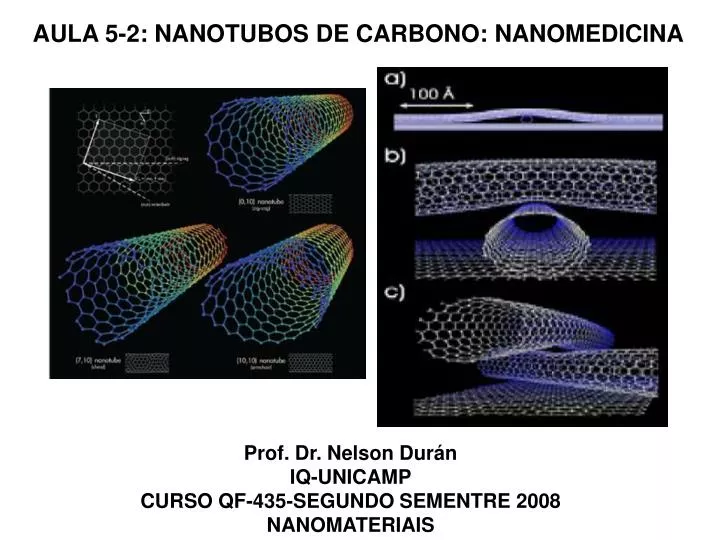
AULA 5-2: NANOTUBOS DE CARBONO: NANOMEDICINA. Prof. Dr. Nelson Durán IQ-UNICAMP CURSO QF-435-SEGUNDO SEMENTRE 2008 NANOMATERIAIS. Bianco, Applications of carbon nanotubes in drug delivery. Current Opinion in Chemical Biology 2005, 9:674–679. human epidermal keratinocytes (HEKs).

E N D
AULA 5-2: NANOTUBOS DE CARBONO: NANOMEDICINA Prof. Dr. Nelson Durán IQ-UNICAMP CURSO QF-435-SEGUNDO SEMENTRE 2008 NANOMATERIAIS
Bianco, Applications of carbon nanotubes in drug delivery. Current Opinion in Chemical Biology 2005, 9:674–679
human epidermal keratinocytes (HEKs) Monteiro-Riviere et al. Surfactant effects on carbon nanotube interactions with human keratinocytes. Nanomedicine: Nanotechnology, Biology, and Medicine 1 (2005) 293– 299
These MWCNTs induced the release of the proinflammatory cytokine interleukin 8 (IL-8)
In conclusion, these studies demonstrate that Pluronic F127 is relatively nontoxic to HEKs in culture but does not increase the cytotoxicity of MWCNTs. In contrast, MWCNTs dosed alone in the aqueous cell culture medium caused significantly more IL-8 release than when MWCNTs were dosed in Pluronic F127. MWCNTs were equally cytotoxic to HEKs with or without the presence of a surfactant. Studies that model nanotube behavior in nonbiological systems suggest that increased dispersion would result in an enhanced ability to interact with cells. Our studies suggest that, although dispersion occurred with surfactant, biological interactions did not correlate with this property.
Tian e al. Cytotoxicity of single-wall carbon nanotubes on human Wbroblasts. Toxicology in Vitro 20 (2006) 1202–1212
Fig. 2. Binding energy intensity of samples with and without purification process: (A) the peaks C 1s and Fe 2p 3/2 in the binding energy intensity are present in SWCNTs before purification; (B) only carbon, peak C1s, is present in purified SWCNTs.
Fig. 3. The effect of refned carbon nanomaterials upon the survival of human fibroblast cells: (A) cells treated with 25 g/ml of CG, MWCNT, CB, AC and SWCNTs, for 1–5 days. Three replicate plates were used for each data point and the experiments were performed at least three times; (B) cells treated with SWCNTs in concentrations of 0.8, 1.61, 3.125, 6.25, 12.5, 25, 50 and 100 g/ml, for 1–5 days.
Fig. 5. Effect of SWCNTs human fibroblast cells: (A) scanning electron microscopy image of dispersed SWCNTs over the substrate, they have the sharpest shape, amid the five nanomaterials, due to a rather large aspect ratio; (B) change in cell spreading seen on samples treated with SWCNTs.
Fig. 6. Morphology of human fibroblast cells observed under transmission electron microscopy images: (A) typical normal cell; (B) cell treated with SWCNTs.
Fig. 7. Phase contrast microscopy images showing the distribution of fibronectin and P-cadherin in normal and SWCNT-treated cells: (A) normal cell; (B) fibronectin (red), P-cadherin (green) and cell nucleus (blue) of a normal cell; (C) SWCNT-treated cell; (D) fibronectin (red), P-cadherin (green) and cell nucleus (blue) of a SWCNT-treated cell.
Fig. 8. Phase contrast microscopy images showing the distribution of FAK protein in normal and SWCNT-treated cells: (A) normal cell; (B) FAK (green) and cell nucleus (blue) in a normal cell; (C) SWCNT-treated cell; (D) FAK (green) and cell nucleus (blue) in a SWCNT-treated cell.
Fig. 9. Phase contrast microscopy images showing the distribution of Factin in normal and SWCNT-treated cells: (A) F-actin (red) and cell nucleus (blue) in a normal cell; (B) F-actin (red) and cell nucleus (blue) in a SWCNT-treated cell.
Their results are twofold. Firstly, it was found that surface area is the variable that best predicts the potential toxicity of these refined carbon nanomaterials, in which SWCNTs induced the strongest cellular apoptosis/necrosis. Secondly, it was found that refined SWCNTs are more toxic than its unrefined counterpart. For comparable small surface areas, dispersed carbon nanomaterials due to a change in surface chemistry, are seen to pose morphological changes and cell detachment, and thereupon apoptosis/necrosis. Finally, it was proposed a mechanism of action that elucidates the higher toxicity of dispersed, hydrophobic nanomaterials of small surface area.
osteosarcoma ROS 17/2.8 cells Zanello et al. Bone Cell Proliferation on Carbon Nanotubes. Nano Lett. 2006, Vol. 6, No. 3 562-567
Figure 3. Morphology of ROS 17/2.8 cells cultured on AP-SWNTs (A-C), AP-MWNTs (D-F), and control cultures on glass cover slips (G-I), as seen with SEM. (A) Osteoblast colony on AP-SWNTs. (B) A flat cell body of a single cell extends over almost the entire field of observation; the cell nucleus protrudes in the center. (C) Tape-like cytoplasmic prolongations (arrow) extend from the flat body of a ROS 17/2.8 cell (a portion of it shown at the left upper corner of the picture) on an evenly distributed AP-SWNT substrate. (D) Osteoblast colony on AP-MWNTs. The nanotubes aggregate unevenly in areas of the glass surface (notice bundles on CNTs on the right). (E) Image of a single ROS 17/2.8 cell on AP-MWNTs. A round single-cell body extends thin neurite-like cytoplasmic prolongations (arrow) that reach the nanotube bundles. (F) SEM micrographs at higher magnification show a detail of long threadlike cytoplasmic prolongations (arrow) that extend from the round body of a single ROS 17/2.8 cell (partially seen on the left, upper corner), interweave with, and reach individual AP-MWNTs. (G) ROS 17/2.8 cell colony cultured on glass. (H) Image of a single cell obtained at higher magnification. (I) Detail of a portion of the cell cytoplasm (covering the left upper half of the picture) in contact with the glass surface; no cytoplasmic prolongations are observed.
As shown in Figure 3A-C, ROS 17/2.8 cell bodies grew flat on AP-SWNTs, similar to their growth on glass (Figure 3G-I). Typical cell diameters for flat ROS 17/2.8 osteoblasts was of the order of 40 ím, which resembled osteoblasts found on the surface of natural bone. However, cell bodies were spherical when cultured on AP-MWNTs. They developed long threadlike cytoplasmic prolongations, as seen in Figure 3D-F. Round cell bodies on MWNTs measured approximately 15 ím in diameter. This cell morphology resembles that of osteocytes, the fully differentiated osteoblasts embedded in the bone matrix. Osteocytes connect and communicate with neighbor cells by means of thin cytoplasmic prolongations that run across the mineralized matrix. Interestingly, a similar neurite-like growth pattern was described for neuronal cultures on AP- and functionalized MWNTs.17 SEM observations performed at high magnification revealed the morphology of physical contacts between cell and CNT materials, as shown in Figure 3C, F, and I. Spherical osteoblasts on AP-MWNTs grew long threadlike cytoplasmic prolongations that reached the nanotube bundles as a way to anchor to a discontinuous, three-dimensional substrate (Figure 3F). These thin pseudopods had diameters in the range of 10-20 nm, close in size to MWNT diameters. Alternatively, flat osteoblasts on AP-SWNTs grew shorter, tape-like cytoplasmic prolongations that adhered to the more evenly distributed layer of nanotubes (Figure 3C). Nanometer-scale cytoplasmic prolongations were not observed in ROS 17/2.8 cells grown on glass (Figure 3I).
It was found that ROS 17/2.8 cells cultured on glass produced cubic crystals after the first week in culture (Figure 4A). These crystals (100-500 nm in length) dispersed at random in intercellular spaces, suggesting that they were not a case of nonspecific ectopic mineralization. On the contrary, ROS 17/2.8 cells cultured on SWNTs produced plate-shaped crystals (100-1000 nm in length, and approximately 20 nm thick) similar in shape to HA crystals found in woven bone, which aggregated in clusters outside the cells (Figure 4B). Although plate-shaped crystals aggregated on the nanomaterials in a disordered fashion, our results indicate that CNTs constitute a suitable substrate for deposition of a mineralized matrix.
Figure 5 shows current-to-voltage relations and raw current traces of a voltage-gated, outward rectifying chloride current that activates at depolarizing potentials in ROS 17/2.8 cells culture. Chloride current amplitude was reduced by 200 M 4,4´-diisothiocyanatostilbene-2,2´-disulfonic acid (DIDS, Sigma), a specific Cl- channel blocker applied to the bath (data not shown). It was not found any statistically significant differences in Cl- current amplitudes and voltage sensitivity in ROS 17/2.8 cells grown on AP-SWNTs and AP-MWNTs. However, current amplitudes measured from cells on AP-MWNTs at membrane potentials over 40 mV were slightly larger than those recorded from AP-SWNT cultures, as shown in the same figure
High-threshold, voltage-activated (HVA) calcium currents were recorded from ROS 17/2.8 cells cultured on AP-SWNTs and AP-MWNTs, as shown in Figure 6. We found that HVA Ca2+ current amplitudes obtained at 20 mV, the membrane voltage value for maximal activation, were ca. twofold larger in osteoblasts cultured on AP-MWNTs than AP-SWNTs. This Ca2+ current was enhanced significantly by 0.5 íM S(-) BayK 8644 (Sigma, Figure 6, left panel), a dihydropyridine agonist specific for L-type Ca2+ channels. Calcium currents were completely blocked by 500 íMCd2+ added to the bath at the end of the experiment (not shown). Inward Ca2+ currents recorded in AP-SWNT and AP-MWNT cultures resembled an L-type Ca2+ channel involved in exocytosis of bone materials described in ROS 17/2.8 cells cultured on plastic dishes. Large Ca2+ current amplitudes obtained in AP-MWNTs correlate with the changes in cell morphology found in this nanomaterial substrate
Normal plasma membrane electrical functions are necessary for osteoblasts to maintain exocytotic activities, and therefore bone formation. Here it was proved that electrical activities of the osteoblast membrane are maintained, and Ca2+ channel functions enhanced, in cells grown on neutral CNTs, confirming a degree of biocompatibility of AP-SWNTs and AP-MWNTs. This is the first description of electrical activities of ion channels in a cell type cultured on CNTs.
Their results provide insight into the understanding of the degree of biocompatibility between live cells and CNTs, and the real possibilities for CNTs to be used as an alternative material for the treatment of bone pathologies that lead to bone loss, with the potential for the regrowth of normal bone. It was found that osteoblasts grow and produce mineralized bone when cultured on electrically neutral CNTs. This growth is diminished by chemical modifications that introduce a net electric charge to the CNTs. In addition, we verified that ROS 17/2.8 cells retain electrical properties necessary for adequate secretion of bone materials when cultured on CNTs. These results suggest that electrically neutral CNTs can be considered as potential filling materials for the treatment of injured bone. It was concluded that CNTs sustain osteoblast growth and bone formation, and thus represent a potential technological advance in the field of bone bioengineering. CNTs show promising biocompatibility with osteoblast cells, and they appear to modulate the cell phenotype. Application of CNTs to bone therapy may lead to the development of new bonegraft materials and techniques.