Download
1 / 9
90 likes | 196 Views
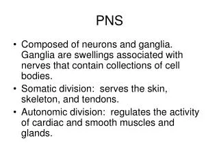
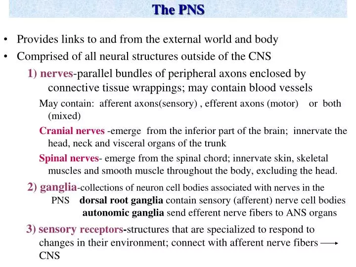
The PNS. Provides links to and from the external world and body Comprised of all neural structures outside of the CNS 1) nerves -parallel bundles of peripheral axons enclosed by connective tissue wrappings; may contain blood vessels

E N D
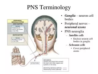
The PNS • Provides links to and from the external world and body • Comprised of all neural structures outside of the CNS 1)nerves-parallel bundles of peripheral axons enclosed by connective tissue wrappings; may contain blood vessels May contain: afferent axons(sensory) , efferent axons (motor) or both (mixed) Cranial nerves -emerge from the inferior part of the brain; innervate the head, neck and visceral organs of the trunk Spinal nerves- emerge from the spinal chord; innervate skin, skeletal muscles and smooth muscle throughout the body, excluding the head. 2) ganglia-collections of neuron cell bodies associated with nerves in the PNS dorsal root ganglia contain sensory (afferent) nerve cell bodies autonomic ganglia send efferent nerve fibers to ANS organs 3)sensory receptors-structures that are specialized to respond to changes in their environment; connect with afferent nerve fibers CNS
The PNS Fig 13.3
The PNS • Comprised of 2 functional divisions: 1) Sensory Division- carries sensory information towardsthe CNS (afferent pathway) • somatic-sensory receptors on skin and skeletal muscle and associated fibers and ganglia • visceral- sensory receptors on glands, smooth muscle, and cardiac muscle and associated fibers and ganglia • sensory ganglia- clusters of cell bodies associated with the somatic and visceral sensory fibers; located in the dorsal root near spinal chord (dorsal root ganglia) • sensory receptors-specialized cellular structures that respond to changes in their environment (stimuli) may be specialized dendrites, neurons or specialized epithelial cells stimulation of sensory receptors results in transient changes in the membrane potential of the sensory membrane (receptor potential) that leads to generation of an afferent impulse (Action Potential) *different from ligand receptors (proteins in cell membranes)
The PNS (con’t) Types of Sensory Receptors: 1) Mechanoreceptors-stimulated by mechanical forces such as touch, stretch (proprioreceptors), vibration 2) Thermoreceptors- stimulated by temperature changes 3)Photoreceptors-stimulated by light 4) Chemoreceptors- respond to chemicals in solution 5) Nociceptors- respond to damaging stimuli that result in pain • Note: other sensory receptor types can result in pain if stimulated by a severe stimulus
The PNS: 2 functional divisions(con’t) 2) Motor Division- carries impulses away from the CNS to affector organs (efferent pathway) somatic-motor neuron fibers that innervate skeletal muscle; cell bodies lie in CNS; neurotransmitter: responsible for voluntary and reflex control of skeletal muscle reflexes - rapid, predictable motor responses to a stimulus function: protection occur over highly specific reflex paths called reflex arcs Components of a reflex arc: 1) sensory receptor stimulus 2) afferent impulse to CNS 3) CNS integration 4) motor neuron activated 5) muscle fiber or organ response
The PNS (con’t) • Components of a spinal reflex arc:
The PNS: 2 functional divisions(con’t) 2) Motor Division- carries impulses away from the CNS to affector organs (efferent pathway) visceral -autonomic (ANS)- motor neuron fibers that innervate glands, smooth muscle and cardiac muscle and associated ganglia neurotransmitters: EPINEPH, NOREPI, ACH • parasympathetic division- active in non-stressful situations; controls body’s maintenance functions (exs:digestion, breathing rate, BP) • sympathetic division- active in stressful situations including vigorous physical activity (exs: pounding heart, sweating, rapid breathing, blood vessel dilation)
The PNS (con’t) Fig. 14.2
The ANS *read pp 525-528 (up to ANS Anatomy) Fig. 14.3