Download
1 / 62
620 likes | 704 Views
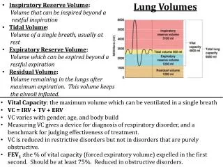
Respiratory Volumes. Tidal volume (TV) – air that moves into and out of the lungs with each breath (approximately 500 ml) Inspiratory reserve volume (IRV) – air that can be inspired forcibly beyond the tidal volume (2100–3200 ml)

E N D
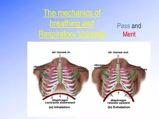
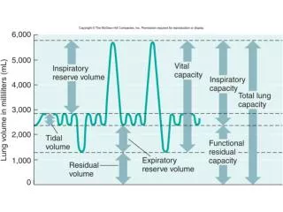
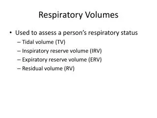
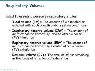
Respiratory Volumes • Tidal volume (TV) – air that moves into and out of the lungs with each breath (approximately 500 ml) • Inspiratory reserve volume (IRV) – air that can be inspired forcibly beyond the tidal volume (2100–3200 ml) • Expiratory reserve volume (ERV) – air that can be evacuated from the lungs after a tidal expiration (1000–1200 ml) • Residual volume (RV) – air left in the lungs after strenuous expiration (1200 ml)
Respiratory Capacities • Inspiratory capacity (IC) – total amount of air that can be inspired after a tidal expiration (IRV + TV) • Functional residual capacity (FRC) – amount of air remaining in the lungs after a tidal expiration (RV + ERV) • Vital capacity (VC) – the total amount of exchangeable air (TV + IRV + ERV) • Total lung capacity (TLC) – sum of all lung volumes (approximately 6000 ml in males); also described as the total amount of gases contained within the lungs
Dead Space • Anatomical dead space – volume of the conducting respiratory passages (150 ml) • Alveolar dead space – alveoli that cease to act in gas exchange due to collapse or obstruction • Total dead space – sum of alveolar and anatomical dead spaces
Pulmonary Function Tests • Spirometer – an instrument consisting of a hollow bell inverted over water, used to evaluate respiratory function • Spirometry can distinguish between: • Obstructive pulmonary disease – increased airway resistance • Restrictive disorders – reduction in total lung capacity from structural or functional lung changes
Pulmonary Function Tests • Total ventilation – total amount of gas flow into or out of the respiratory tract in one minute • Forced vital capacity (FVC) – gas forcibly expelled after taking a deep breath • Forced expiratory volume (FEV) – the amount of gas expelled during specific time intervals of the FVC
Pulmonary Function Tests • Increases in TLC (total lung capacity), FRC (functional residual capacity), and RV (residual volume) may occur as a result of obstructive disease • Reduction in VC, TLC, FRC, and RV result from restrictive disease
Alveolar Ventilation • Alveolar ventilation rate (AVR) – measures the flow of fresh gases into and out of the alveoli during a particular time, typically 1 minute • Slow, deep breathing increases AVR and rapid, shallow breathing decreases AVR
Nonrespiratory Air Movements • Most result from reflex action • Are used to clear airways, express emotions, speak, etc. • Examples include: coughing, sneezing, crying, laughing, hiccupping, and yawning
Basic Properties of Gases: Dalton’s Law of Partial Pressures • Total pressure exerted by a mixture of gases is the sum of the pressures exerted independently by each gas in the mixture • The partial pressure of each gas is directly proportional to its percentage in the mixture
Basic Properties of Gases: Henry’s Law • When a mixture of gases is in contact with a liquid, each gas will dissolve in the liquid in proportion to its partial pressure • The amount of gas that will dissolve in a liquid also depends upon its solubility: • Carbon dioxide is the most soluble • Oxygen is 1/20th as soluble as carbon dioxide • Nitrogen is practically insoluble in plasma
Composition of Alveolar Gas • The atmosphere is mostly oxygen and nitrogen, while alveoli contain more carbon dioxide and water vapor • These differences result from: • Gas exchanges in the lungs – oxygen diffuses from the alveoli and carbon dioxide diffuses into the alveoli • Humidification of air by conducting passages • The mixing of alveolar gas that occurs with each breath
External Respiration: Pulmonary Gas Exchange • Factors influencing the movement of oxygen and carbon dioxide across the respiratory membrane • Partial pressure gradients and gas solubilities • Matching of alveolar ventilation and pulmonary blood perfusion • Structural characteristics of the respiratory membrane
Partial Pressure Gradients and Gas Solubilities • The partial pressure oxygen (PO2) of venous blood is 40 mm Hg; the partial pressure in the alveoli is 104 mm Hg • This steep gradient shows that oxygen partial pressures reaches equilibrium (high enough pressure is achieved that O2 can move into the blood vessels) rapidly, and thus blood can move very rapidly through the pulmonary capillary and still be adequately oxygenated
Partial Pressure Gradients and Gas Solubilities • Although carbon dioxide has a lower partial pressure gradient: • It is 20 times more soluble in plasma than oxygen • It diffuses in equal amounts with oxygen
Ventilation-Perfusion Coupling • Ventilation – the amount of gas reaching the alveoli • Perfusion – the blood flow reaching the alveoli • Ventilation and perfusion must be tightly regulated for efficient gas exchange
Ventilation-Perfusion Coupling • Changes in PCO2 (partial pressure of CO2) in the alveoli cause changes in the diameters of the bronchioles • Passageways servicing areas where alveolar carbon dioxide is high dilate • Those serving areas where alveolar carbon dioxide is low constrict
Surface Area and Thickness of the Respiratory Membrane • Respiratory membranes: • Are only 0.5 to 1 m thick, allowing for efficient gas exchange • Have a total surface area (in males) of about 60 m2 (40 times that of one’s skin) • Thicken if lungs become waterlogged and edematous, whereby gas exchange is inadequate and oxygen deprivation results • Decrease in surface area with emphysema, when walls of adjacent alveoli break through
Internal Respiration • The factors promoting gas exchange between systemic capillaries and tissue cells are the same as those acting in the lungs • The partial pressures and diffusion gradients are reversed • PO2 in tissue is always lower than in systemic arterial blood • PO2 of venous blood draining tissues is 40 mm Hg and PCO2 is 45 mm Hg
Oxygen Transport • Molecular oxygen is carried in the blood: • Bound to hemoglobin (Hb) within red blood cells • Dissolved in plasma
Oxygen Transport: Role of Hemoglobin • Each Hb molecule binds four oxygen atoms in a rapid and reversible process • The hemoglobin-oxygen combination is called oxyhemoglobin (HbO2) • Hemoglobin that has released oxygen is called reduced hemoglobin (HHb) Lungs HHb + O2 HbO2 + H+ Tissues
Hemoglobin (Hb) • Saturated hemoglobin – when all four hemes of the molecule are bound to oxygen • Partially saturated hemoglobin – when one to three hemes are bound to oxygen • The rate that hemoglobin binds and releases oxygen is regulated by: • PO2, temperature, blood pH, PCO2, and the concentration of BPG (an organic chemical) • These factors ensure adequate delivery of oxygen to tissue cells
Influence of PO2 on Hemoglobin Saturation • 98% saturated arterial blood contains 20 ml oxygen per 100 ml blood (20 vol %) • As arterial blood flows through capillaries, 5 ml oxygen are released • The saturation of hemoglobin in arterial blood explains why breathing deeply increases the PO2 but has little effect on oxygen saturation in hemoglobin
Hemoglobin Saturation Curve • Hemoglobin is almost completely saturated at a PO2 of 70 mm Hg • Further increases in PO2 produce only small increases in oxygen binding • Oxygen loading and delivery to tissue is adequate when PO2 is below normal levels
Hemoglobin Saturation Curve • Only 20–25% of bound oxygen is unloaded during one systemic circulation • If oxygen levels in tissues drop: • More oxygen dissociates from hemoglobin and is used by cells • Respiratory rate or cardiac output need not increase
Other Factors Influencing Hemoglobin Saturation • Temperature, H+, PCO2, and BPG • Modify the structure of hemoglobin and alter its affinity for oxygen • Increases of these factors: • Decrease hemoglobin’s affinity for oxygen • Enhance oxygen unloading from the blood • Decreases act in the opposite manner • These parameters are all high in systemic capillaries where oxygen unloading is the goal
Factors That Increase Release of Oxygen by Hemoglobin • As cells metabolize glucose, carbon dioxide is released into the blood causing: • Increases in PCO2 and H+ concentration in capillary blood • Declining pH (acidosis), which weakens the hemoglobin-oxygen bond (Bohr effect) • Metabolizing cells have heat as a byproduct and the rise in temperature increases BPG synthesis • All these factors ensure oxygen unloading in the vicinity of working tissue cells
Hemoglobin-Nitric Oxide Partnership • Nitric oxide (NO) is a vasodilator that plays a role in blood pressure regulation • NO is free radical, a by-product of combustion (as from automobiles) and has a very short half life (a few seconds) in the blood • Hemoglobin is a vasoconstrictor and a nitric oxide scavenger (heme destroys NO) • However, as oxygen binds to hemoglobin: • Nitric oxide binds to a amino acid on hemoglobin (cysteine) • Bound nitric oxide is protected from degradation by hemoglobin’s iron
Hemoglobin-Nitric Oxide Partnership • The hemoglobin is released as oxygen is unloaded, causing vasodilation • As deoxygenated hemoglobin picks up carbon dioxide, it also binds nitric oxide and carries these gases to the lungs for unloading
Carbon Dioxide Transport • Carbon dioxide is transported in the blood in three forms • Dissolved in plasma – 7 to 10% • Chemically bound to hemoglobin – 20% is carried in RBCs as carbaminohemoglobin • Bicarbonate ion in plasma – 70% is transported as bicarbonate (HCO3–)
Transport and Exchange of Carbon Dioxide • Carbon dioxide diffuses into RBCs and combines with water to form carbonic acid (H2CO3), which quickly dissociates into hydrogen ions and bicarbonate ions • In RBCs, carbonic anhydrase reversibly catalyzes the conversion of carbon dioxide and water to carbonic acid
Transport and Exchange of Carbon Dioxide • At the tissues: • Bicarbonate quickly diffuses from RBCs into the plasma • The chloride shift – to counterbalance the outrush of negative bicarbonate ions from the RBCs, chloride ions (Cl–) move from the plasma into the erythrocytes
Transport and Exchange of Carbon Dioxide • At the lungs, these processes are reversed • Bicarbonate ions move into the RBCs and bind with hydrogen ions to form carbonic acid • Carbonic acid is then split by carbonic anhydrase to release carbon dioxide and water • Carbon dioxide then diffuses from the blood into the alveoli
Haldane Effect • The amount of carbon dioxide transported is markedly affected by the PO2 • Haldane effect – the lower the PO2 and hemoglobin saturation with oxygen, the more carbon dioxide can be carried in the blood
Haldane Effect • At the tissues, as more carbon dioxide enters the blood: • More oxygen dissociates from hemoglobin (Bohr effect) • More carbon dioxide combines with hemoglobin, and more bicarbonate ions are formed • This situation is reversed in pulmonary circulation
Influence of Carbon Dioxide on Blood pH • The carbonic acid–bicarbonate buffer system resists blood pH changes • If hydrogen ion concentrations in blood begin to rise, excess H+ is removed by combining with HCO3– • If hydrogen ion concentrations begin to drop, carbonic acid dissociates, releasing H+
Influence of Carbon Dioxide on Blood pH • Changes in respiratory rate can also: • Alter blood pH • Provide a fast-acting system to adjust pH when it is disturbed by metabolic factors
Control of Respiration: Medullary Respiratory Centers • The dorsal respiratory group (DRG), or inspiratory center: • Is located near the root of nerve IX • Appears to be the pacesetting respiratory center • Excites the inspiratory muscles and sets eupnea (12-18 breaths/minute) • Becomes dormant during expiration • The ventral respiratory group (VRG) is involved in forced inspiration and expiration
Control of Respiration: Pons Respiratory Centers • Pons centers: • Influence and modify activity of the medullary centers • Smooth out inspiration and expiration transitions and vice versa • The pontine respiratory group (PRG) – continuously inhibits the inspiration center, cyclically, by limiting the action potentials of the phrenic nerve
Respiratory Rhythm • A result of reciprocal inhibition of the interconnected neuronal networks in the medulla • Other theories include • Inspiratory neurons are pacemakers and have intrinsic automaticity and rhythmicity • Stretch receptors in the lungs may help establish respiratory rhythm
Depth and Rate of Breathing • Inspiratory depth is determined by how actively the respiratory center stimulates the respiratory muscles • Rate of respiration is determined by how long the inspiratory center is active • Respiratory centers in the pons and medulla are sensitive to both excitatory and inhibitory stimuli
Depth and Rate of Breathing: Reflexes • Pulmonary irritant reflexes – irritants promote reflexive constriction of air passages • Inflation reflex (Hering-Breuer) – stretch receptors in the lungs are stimulated by lung inflation • Upon inflation, inhibitory signals are sent to the medullary inspiration center to end inhalation and allow expiration
Depth and Rate of Breathing: Higher Brain Centers • Hypothalamic controls act through the limbic system to modify rate and depth of respiration • Example: breath holding that occurs in anger • A rise in body temperature acts to increase respiratory rate • Cortical controls are direct signals from the cerebral motor cortex that bypass medullary controls • Examples: voluntary breath holding, taking a deep breath
Depth and Rate of Breathing: PCO2 • Changing PCO2 levels are monitored by chemoreceptors of the brain stem • Carbon dioxide in the blood diffuses into the cerebrospinal fluid where it is hydrated • Resulting carbonic acid dissociates, releasing hydrogen ions • PCO2 levels rise (hypercapnia) resulting in increased depth and rate of breathing
Depth and Rate of Breathing: PCO2 • Hyperventilation – increased depth and rate of breathing that: • Quickly flushes carbon dioxide from the blood • Occurs in response to hypercapnia (too much CO2 in the blood stream) or emotional distress • Though a rise CO2 acts as the original stimulus, control of breathing at rest is regulated by the hydrogen ion concentration in the brain
Depth and Rate of Breathing: PCO2 • Hypoventilation – slow and shallow breathing due to abnormally low PCO2 levels • Apnea (breathing cessation) may occur until PCO2 levels rise
Depth and Rate of Breathing: PCO2 • Arterial oxygen levels are monitored by the aortic and carotid bodies • Substantial drops in arterial PO2 (to 60 mm Hg) are needed before oxygen levels become a major stimulus for increased ventilation • If carbon dioxide is not removed (e.g., as in emphysema and chronic bronchitis), chemoreceptors become unresponsive to PCO2 chemical stimuli • In such cases, PO2 levels become the principal respiratory stimulus (hypoxic drive)
Depth and Rate of Breathing: Arterial pH • Changes in arterial pH can modify respiratory rate even if carbon dioxide and oxygen levels are normal • Increased ventilation in response to falling pH is mediated by peripheral chemoreceptors
Depth and Rate of Breathing: Arterial pH • Acidosis may reflect: • Carbon dioxide retention • Accumulation of lactic acid • Excess fatty acids in patients with diabetes mellitus • Respiratory system controls will attempt to raise the pH by increasing respiratory rate and depth
Respiratory Adjustments: Exercise • Respiratory adjustments are geared to both the intensity and duration of exercise • During vigorous exercise: • Ventilation can increase 20 fold • Breathing becomes deeper and more vigorous, but respiratory rate may not be significantly changed (hyperpnea) • Exercise-enhanced breathing is not prompted by an increase in PCO2 or a decrease in PO2 or pH • These levels remain surprisingly constant during exercise