Download
1 / 61
640 likes | 929 Views
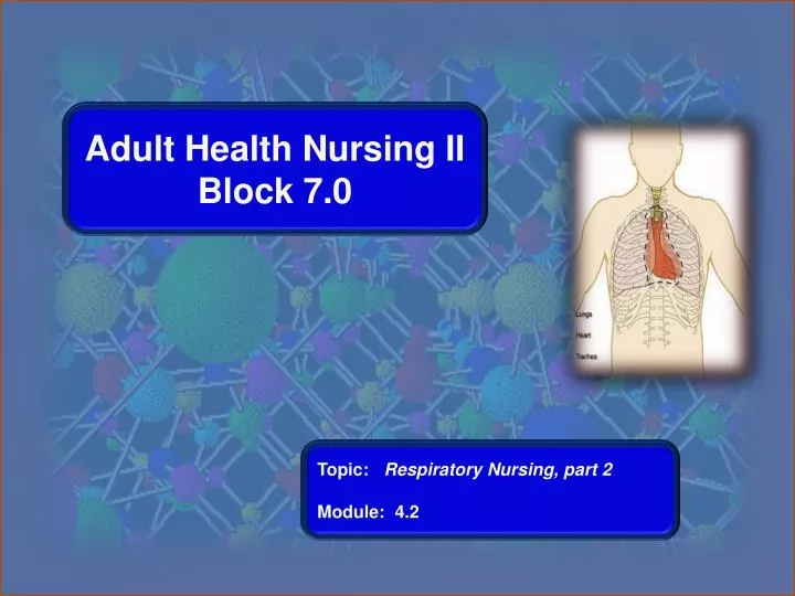
Adult Health Nursing II Block 7.0. Topic: Respiratory Nursing, part 2 Module : 4.2. Lung Cancer. Cause: chronic tissue irritation or inflammation d/t repeated exposure to inhaled substances (cigarette smoke, occupational or environmental agents)

E N D
Adult Health Nursing II Block 7.0 Topic: Respiratory Nursing, part 2 Module: 4.2
Lung Cancer • Cause: chronic tissue irritation or inflammation d/t repeated exposure to inhaled substances (cigarette smoke, occupational or environmental agents) • 80-90% linked to cigarette smoking (includes 2nd-hand smoke) Block 7.0 Module 4.1
Lung Cancer • Leading cause of cancer deaths in both men & women accounting for 28% of all cancer deaths (>165,000 deaths/year) • 5-year survival rate only 14% • Slow growing – takes 8-10 yr to reach 1cm, smallest detectable lesion on an x-ray • Low survival rate d/t dx at a late state when metastasis (spread) has already occurred • Metastasize by (1) direct extension; (2) thru the blood (hematogenous); & invading lymph glands & vessels. Block 7.0 Module 4.1
S/Sx of Lung Cancer • Insidious, often nonspecific, appearing late in disease process • #1 sx: dry, persistent cough or change to chronic, productive cough • Hemoptysis (coughing up blood) • Recurrent lung infections w/chills, fever • Dyspnea; painful breathing; wheezing • Weight loss, fatigue Block 7.0 Module 4.1
Diagnostic & Lab Tests • Chest x-ray, chest CT • Sputum cytology • Bronchoscopy/mediastinoscopy w/biopsy • Needle biopsy • MRI • PET scan to detect metastasis • CEA (carcinoembryonic antigen titer) Block 7.0 Module 4.1
Medical Management • May include combination of surgery, chemo, & radiation therapies • Chemotherapy may provide pain relief but does not usually cure • Useful in rx of mets to brain, spine, pericardium • Side effects: N/V, alopecia (hair loss), anemia, immunosuppression, mouth sores thrombocytopenia (decreased platelets) • Radiation therapy may cure, relieve sx, reduce size of tumor Block 7.0 Module 4.1
Surgical Management • Preferred rx, esp. if non-small cell CA & no mets • Lobectomy – resection of entire lobe • Pneumonectomy – resection of entire lung • Segmentectomy – resection of bronchus, pulmonary artery & vein, & portion of involved lung segment • Wedge resection – removal of peripheral portion of small, local areas Block 7.0 Module 4.1
Interventions for Palliation • Oxygen therapy • Drug therapy • Radiation therapy • Laser therapy • Thoracentesis and pleurodesis • Dyspnea management • Pain management • Hospice & end-of-life issues Block 7.0 Module 4.1
Nursing Responsibilities • Manage pain, n/v, dyspnea, fatigue • Drugs for sx relief • Oxygen • Ways to reduce fatigue • Psychological support for pt & family • Identify community resources • Help family deal with poor prognosis • End-of-life treatment options (hospice, home health) Block 7.0 Module 4.1
Pulmonary Edema Pulmonary edema is swelling and fluid accumulation in the lungs. The extra fluid and swelling drown the patient by impairing healthy gas exchange with the circulating blood and can cause respiratory failure. Block 7.0 Module 4.1
Treatment for Block 7.0 Module 4.1
Pulmonary Embolism (PE) • Clot enters bloodstream & lodges in pulmonary vessels. • Blood clot is most common, but may also be fat, air, amniotic fluid, tumor tissue. • Obstructs pulmonary blood flow, leading to decreased systemic oxygenation, pulmonary tissue hypoxia & potential death. • 90-95% of PE arise from DVTs (deep vein thrombosis) in the leg. • 10% mortality rate; many die within 1st hour Block 7.0 Module 4.1
Pulmonary Embolus (PE) Block 7.0 Module 4.1
Risk Factors for PE • DVT #1 90-95% • Prolonged immobility (lying or sitting) • Central venous catheters, including PICCs • Surgery (orthopedic, pelvic, abdominal, recent pregnancy/childbirth) • Obesity • Advanced age • Hypercoagulability (anemia, estrogen therapy, birth control pills, smoking) • History of thromboembolism Block 7.0 Module 4.1
Symptoms (subjective): Dyspnea, sudden onset Sharp, inspiratory chest pain Apprehension, restlessness Feeling of impending doom Signs (objective): Tachypnea, gasping Crackles, diminished breath sounds Cough, hemoptysis Tachycardia Hypotension Fever, low grade Decreased SaO2 S/Sx of PE Block 7.0 Module 4.1
Diagnostic & Lab Tests • Spiral CT most often used to dx PE • ABGs – indicate hypoxemia, hypocapnia initially (respiratory alkalosis) later will have hypercarbia w/respiratory acidosis mixed w/metabolic acidosis d/t lactic acid buildup • Venous U/S to determine presence of DVT to support PE dx • Pulmonary angiogram is most specific test but not usually done d/t risk Block 7.0 Module 4.1
Pharmacology for PE • Heparin (an anticoagulant) is initial treatment of choice • Keeps embolus from enlarging & prevents formation of new clots. Does not dissolve clot. Pt’s own body dissolves the clot. • High risk for bleeding. • Monitor lab: therapeutic range for PTT/aPTT is 1.5-2 x baseline (baseline usually 25-39 sec) (see sample heparin protocol sheet) (see Chart 34-5, p. 682) • Antidote for heparin overdose: protamine sulfate IV • Avoid antiplatelet drugs like aspirin & Plavix increases risk of bleeding Block 7.0 Module 4.1
Pharmacology for PE • Warfarin (Coumadin) (an anticoagulant) is started on day 3 of heparin therapy long half-life (3-5 days) • Pt continues on both heparin & warfarin until INR 2-3, then heparin d/c’d. • Monitor lab: Therapeutic range for INR: 2-3 • Antidote for coumadin overdose: Vit. K SQ or IV • Avoid aspirin & acetaminaphen (increases risk for bleeding) • Avoid foods high in Vit K (green, leafy vegetables decrease effects of warfarin) Block 7.0 Module 4.1
Pharmacology for PE • Streptokinase (a thrombolytic/fibrinolytic drug) – used in massive PE with shock &/or hypotension to dissolve clot. HIGH risk for bleeding. Bleeding is most common side effect. • Other anticoagulants – LMWH (low molecular weight heparin) – Lovenox SQ 1mg/kg • Pain meds, antianxiety meds Block 7.0 Module 4.1
Interventions for PE • O2 • Monitor q1-2 hr & prn: • Vital signs • Respiratory status (lung sounds, crackles, cyanosis, increased dyspnea) • C/V status (dysrhythmias, edema) • Surgery -- Embolectomy if clot is very large & if fibrinolytic therapy contraindicated (hx of cerebral or GI bleed) -- Inferior vena cava filter (Greenfield filter) placement in high risk patients, esp. if anticoagulants are contraindicated Block 7.0 Module 4.1
Nursing Interventions for PE • Bedrest (24-48 hr) in semi-Fowler’s position • Turn, cough & deep breath • O2: monitor ABGs, SaO2 , nebulizer rx, incentive spirometer • Monitor q1-2h & prn: vital signs, respiratory status (lung sounds, crackles, cyanosis, increased dyspnea), & C/V status (edema, dysthythmias, chest pain) • Assess for internal & external bleeding • Assess for +Homans’ sign (unreliable) • Assess for s/sx of obvious &/or occult bleeding (easy bruising, blood in stools/urine/emesis) • See Chart 34-6, p. 683 Block 7.0 Module 4.1
Homan’s Sign Forced plantar flexion of the ankle may elicit pain response in leg. Unreliable do not use. Block 7.0 Module 4.1
Health Promotion & Prevention of PE • Stop smoking esp. if on birth control pills • Reduce weight, increase physical activity • Anticoagulants for pts w/atrial fib • Anticoagulants & compression stockings for post-op & other at-risk pts • Ambulate pt ASAP post-op • If traveling or sitting for long periods, get up frequently & drink plenty of fluids. • Refrain from massaging leg muscles. • Avoid tight garters, girdles, belts • Prevent pressure under the popliteal space (don’t put pillows under pt’s knees) Block 7.0 Module 4.1
Patient Education for Anticoagulants • Prevent bleeding from anticoagulants • Use electric razor • Avoid sharps • Soft bristle toothbrush • No OTC meds w/o MD’s permission • Avoid laxatives, may affect Vit K absorption • Report dark, tarry stools • Wear ID or carry med card Block 7.0 Module 4.1
Chest Trauma • About 25% of civilian traumatic deaths result from chest injuries • Blunt chest trauma: sudden pressure to chest wall. Most common: • Steering wheel or seatbelt in MVA • Fall • Bicycle crash • Penetrating trauma: foreign object penetrates chest wall. Most common: • Stabbing • Gunshot wounds Block 7.0 Module 4.1
Assessment & Diagnostics for Chest Trauma • Assess for patent airway • Assess for bleeding, open wounds • Assess rate, depth, symmetry of resp • Assess for stridor (late sign), cyanosis, trauma to mouth, face, neck • Assess VS & neuro status • CXR, CT, CBC, lytes, ABGs, SaO2, EKG • Totally undress pt so nothing is missed Block 7.0 Module 4.1
Pulmonary Contusion • Most common chest injury in U.S. • Often results from rapid deceleration in MVA • Respiratory failure develops over time rather than immediately • Damage to lung tissues resulting in hemorrhage & localized edema decreased lung movement & gas exchange • May not be initially evident (even on CXR), may not develop until 1-2 days post injury • S/sx: dyspnea, hemoptysis, hypoxia • Rx: O2 support, analgesics (opioids), ATBs, may need mechanical vent if ARDS Block 7.0 Module 4.1
Rib Fractures • Rib fractures 2nd most common chest injury, usually d/t blunt trauma • Uncomplicated rib fx heal spontaneously • S/sx: severe chest pain resulting in compromised respirations; possible crepitus if rib punctures lung • Main focus: pain control so pt’s respirations will not be compromised • Avoid analgesics that cause respiratory depression Block 7.0 Module 4.1
Flail Chest • Caused by multiple rib fractures resulting in instability of chest wall with paradoxical breathing – portion of lung under injured chest wall moves in on inspiration & out on expiration • Usually unilateral • Results in severe respiratory distress w/decreased gas exchange & ability to cough • High mortality (40%), esp. in older pts • S/sx: pain, dyspnea, cyanosis, SOB, tachycardia, hypotension, anxiety Block 7.0 Module 4.1
Flail Chest Block 7.0 Module 4.1
Interventions for Flail Chest • Maintain patent airway • Agitation, irrational, combative behavior may indicate decreased O2 to the brain • Maintain fluid volume • Maintain chest wall integrity • Stabilized w/positive-pressure ventilation Block 7.0 Module 4.1
Interventions for Flail Chest • Humidified O2 • Analgesics (opioids) • Turn, cough, deep breath • May need mechanical vent if shock or respiratory failure occurs • Monitor: ABGs, VS, fluid & electrolyte balance for hypovolemia or shock Block 7.0 Module 4.1
Problems of the Pleural Space • Lies between the parietal pleura (membrane lining the chest cavity) and the visceral pleura (surrounds the lungs) • Holds about 50 ml of lubricating fluid • Creates a negative pressure that keeps the lungs expanded • Excess fluid or air accumulation in the pleural space limits lung expansion and leads to respiratory distress Block 7.0 Module 4.1
Pleural Space Block 7.0 Module 4.1
PROBLEMS OF THE PLEURA • Pneumothorax: air in pleural space • Hemothorax: blood in pleural space • Pleural effusion: fluid in pleural space • Pulmonary Empyema: pus in pleural space • Pleurisy: inflammation of the pleura Block 7.0 Module 4.1
Pneumothorax &/or Hemothorax • Pneumothorax: Air enters pleural space • Hemothorax: Blood enters pleural space • Prevents lung expansion & exchange of O2 & CO2. • Causes the lung to collapse • Severity depends on amount of lung that is collapsed Block 7.0 Module 4.1
Pneumothorax &/or Hemothorax Block 7.0 Module 4.1
S/sx of Pneumothorax/Hemothorax • Sudden onset of pleuritic pain • Tachypnea, dyspnea • Anxiety, apprehension • Reduced or absent breath sounds on affected side • Hypotension, tachycardia • Crepitus (subcutaneous emphysema) Block 7.0 Module 4.1
Causes for Pneumo/Hemothorax • Open pneumothorax: sharp chest wound (stab or gunshot wound, surgical thoracotomy, thoracentesis, chest tube placement, lung biopsy) • Closed pneumothorax: no external wound • Interstitial lung disease (cancer, TB) • ARDS • Mechanical ventilation Block 7.0 Module 4.1
Interventions for Pneumo/Hemothorax • Goal: evacuation of air &/or blood from pleural space • Oxygen therapy • Pain management • Thoracentesis • Chest tube to water seal and/or suction • Patient with hemothorax may need open thoracotomy for massive (>1500 mL) &/or persistent bleed (>200 mL over 3 hours) • Monitor: VS, respiratory status, blood loss, chest tubes Block 7.0 Module 4.1
PLEURODESIS Procedure that causes the pleura around the lung to stick together and prevents the buildup of fluid in the pleural space. This procedure is done in cases of severe recurrent pleural effusion (fluid around the lungs), as from cancer, to prevent the reaccumulation of fluid. In pleurodesis, an irritant (such as sterile talc powder) is instilled inside the space between the pleura in order to create inflammation which tacks the two pleura together. This procedure obliterates the space between the pleura and prevents re-accumulation of fluid. Block 7.0 Module 4.1
Pleurodesis Block 7.0 Module 4.1
Tension Pneumothorax • Collapse of lung d/t air entering the pleural space on inspiration, but does not leave on expiration heart, great vessels & thorax in mediastinum shifts to unaffected side • Pressure in lung decreases venous return leading to decreased filling of the heart & decreased cardiac output. • Develops rapidly, quickly fatal if not detected & treated Block 7.0 Module 4.1