Download
1 / 21
210 likes | 223 Views
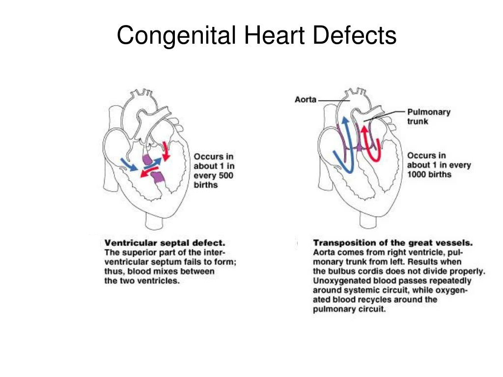
Congenital Heart Defects. Congenital Heart Defects. Congenital Heart Defects. Electrical Conduction System. Sino Atrial (SA) Node – sup/post rt atrium. Atrial Ventricular (AV) Node – inf rt atrium. AV Bundle (of His) – sup IV septum. L and R Bundle Branches – IV septum.

E N D
Electrical Conduction System Sino Atrial (SA) Node – sup/post rt atrium Atrial Ventricular (AV) Node – inf rt atrium AV Bundle (of His) – sup IV septum L and R Bundle Branches – IV septum Purkinje Fibers – both ventricles
The Electrocardiogram Recording of the electrical activities in the heart P wave = Atrial Depolarization QRS complex = Ventricular Depolarization T wave = Ventricular Repolarization
Blood = Plasma + Formed (Cellular) Elements Plasma • ~ 55% blood volume • ~ 92% of plasma is water • High dissolved O2 content • Dissolved proteins • Minerals, glucose, ions. Cells • ~ 45% blood volume • RBCs (~ 99% of cells) • WBCs (~ 1% of cells)
The Proteins in Plasma • Albumins • 60% of plasma proteins. • Globulins • 35% of plasma proteins. • Fibrinogen • For clotting reaction, forms fibrin. • * serum = plasma without clotting proteins
Cellular Components RBCs (erythrocytes) ~ 99% of all cells. Hematocrit = % of blood occupied by cellular components. (packed RBC volume) Lacks nuclei, mitochondria and ribosomes. Anaerobic metabolism Life span = ~120 days
Scanning Electron Micrograph (SEM) of Erythrocytes or Red Blood Cells (RBCs) on the tip of a hypodermic needle.
White Blood Cells (Leukocytes) Granular Leukocytes • Neutrophils 70% of circulating leukocytes • Multi-lobed nucleus (3 or more), mobile phagocytes. • Eosinophils much less common • Bi-lobed nucleus with ‘orange’ staining granules. • Basophils relatively rare • Bi-lobed with dark staining granules, releases histamine.
Agranular Leukocytes • Lymphocytesprimary cell of lymphatic system • T-cells attack foreign cells directly. • B-cells produce antibodies. • Monocytes • Large nucleus, differentiate intoMacrophages.
n l e
Platelet cells (Thrombocytes) • Fragments of cells (Megakaryocytes) for clotting. Never Let Monkeys Eat Bananas
Arteries Arterioles Capillaries Heart! Venules Veins
Control of the Heart • Basic rate established by pacemaker cells inside the heart (myocardium) – called “intrinsic myogenic control” • Modified by Autonomic N.S. (ANS) • Para: decreases rate via the Vagus n. X. • Sym: increases heart rate and force of contraction via cardiac accelerator n.