Download
1 / 77
850 likes | 1.47k Views
TRIGEMINAL NEURALGIA. CONTENTS. INTRODUCTION ETIOPATHOGENESIS DIAGNOSIS MEDICINAL MANAGEMENT SURGICAL MANAGEMENT. Introduction. Disorder characterized by lancinating attacks of severe facial pain

E N D
CONTENTS • INTRODUCTION • ETIOPATHOGENESIS • DIAGNOSIS • MEDICINAL MANAGEMENT • SURGICAL MANAGEMENT
Introduction • Disorder characterized by lancinating attacks of severe facial pain • Diagnosis based primarily on a history of characteristic pain attacks that are consistent with specific research & clinical criteria
In majority of patients, clinical exam-ination, imaging and lab tests are unremarkable – Classic TN • In a smaller group, signs & symptoms secondary to another disease affecting the trigeminal system – Symptomatic TN
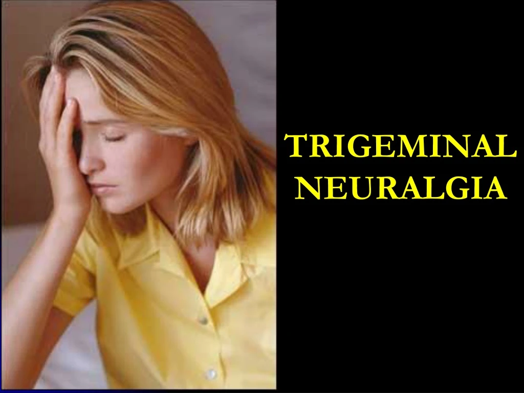
Definition Trigeminal neuralgia is defined as Pain in the distribution of one or more branches of the 5th cranial nerve sudden Usually unilateral Severe Brief Stabbing Lancinating Recurring
Epidemiology and Demographics - Incidence of approx 4 in 100,000 Familial cases also reported • Majority of cases occur spontaneously • Slight female predominance • Over age 50
Pain typically consists of lancinatingparoxysms • Mostly in Second & Third trigeminal divisions • Right side most often involved • Pain attacks stereotyped • Symptomfree between attacks • Chronic disorder, most patients will experience pain attacks for years unless appropriately treated
Etiology and Pathogenesis Cause – not known Injury to the nerve root – an initiating factor? (Benign tumors and vascular anomalies that compress the trigeminal nerve root can produce symptoms clinically indistinguishable from classic TN)
Based on the morphologic and physio-logic changes following partial nerve injury, Devor et al proposed “ignition hypothesis”. A trigeminal injury induces physiologic changes that result in a population of hyper-excitable and functionally linked primary sensory neurons. The discharge of any individual neuron of this group can quickly spread to activate the entire population. Such a discharge could underlie the sudden jolt of pain in TN attack.
Diagnosis • Clinical • Consider in all patients with unilateral facial pain • Prompt Diagnosis important as pain can be severe • Distinguish classical from symptomatic for treatment purposes • Look for “red flags” of other diseases
Red Flags • Abnormal Neuro exam • Abnormal oral, dental, or ear exam • Age < 40 yrs • Bilateral Symptoms • Dizziness or vertigo
Red Flags • Hearing loss • Numbness • Pain lasting > 2 minutes • Pain outside of trigeminal distribution • Visual changes
Diagnostic History • Very important • Recurrent, unilateral facial pain • Lasts seconds • May recur 100’s of times per day • Pain : • Severe Stereotypical • Sharp Stabbing • Superficial Shock-like
Diagnostic History • 1 or more of the nerve’s divisions • Trigger factors: • Talking Shaving • Smiling Applying make-up • Chewing Wind • Teeth brushing • Age > 40 yrs. • Asymptomatic time or not ?
Physical Exam • Usually a normal exam • Useful for identifying abnormals that point to other Diagnoses. • HEENT, including TMJ & Masseter • Oral exam, including teeth & gums • Neuro exam • Check for trigger zones
Clinical Presentation andPhysical Findings Diagnosis of TN based on distinctive signs & symptoms. White & Sweet articulated diagnostic criteria for TN. Consists of 5 major clinical features that define the diagnosis of TN
Sweet diagnostic criteria • Pain is paroxysmal • The pain may be provoked by light touch to the face (trigger zones) • The pain is confined to the trigeminal distribution • The pain is unilateral • The clinical sensory examination is normal
Patients who did not meet all the criteria rarely benefited. The Sweet criteria were incorporated into the criteria published by IASP & IHS. ICHDII(IHS)subdivides Trigeminal Neuralgia (code 13.1) into, - Classic TN (code 13.1.1) - Symptomatic TN (code 13.1.2)
Classic TN (13.1.1) Most common form- idiopathic, and also associated with vascular compression. “a unilateral disorder characterized by brief electric shock-like pains, abrupt in onset and termination, limited to the distribution of one or more divisions of trigeminal nerve. Pain is commonly evoked by trivial stimuli including washing, shaving, smoking, talking and/or brushing the teeth (trigger factors) and frequently occurs spontaneously. Small areas in the nasolabial fold and/or chin may be particularly susceptible to the precipitation of pain (trigger areas). The pains usually remit for variable periods.”
ICHD Criteria for Classical TN (13.1.1) • Paroxysmal attacks of pain lasting from a fraction of a second to 2 minutes, affecting one or more divisions of the trigeminal nerve and fulfilling criteria B and C • Pain has at least one of the following characteristics: 1. intense, sharp, superficial or stabbing 2. precipitated from trigger areas or by trigger factors • Attacks are stereotyped in individual patient. • There is no clinically evident neurological deficit. • Not attribute to another disorder.
Symptomatic TN (13.1.2) - Results from another disease process (MS or a cerebellopontine angle tumor) “Pain indistinguishable from 13.1.1 classic TN but caused by a demonstrable structural lesion other than vascular compression.”
ICHD Criteria for Symptomatic TN (13.1.2) • Paroxysmal attacks of pain lasting from a fraction of a second to 2 minutes, with or without persistence of aching between paroxysms, affecting one or more divisions of trigeminal nerve and fulfilling criteria B and C. • Pain has at least one of the following characteristics: 1. Intense, sharp, superficial or stabbing 2. Precipitated from trigger areas or by trigger factors. • Attacks are stereotyped in individual patient. • A causative lesion, other than vascular compression, has been demonstrated by special investigations and/or posterior fossa exploration.
The pain of TN…… • Paroxysmal attacks • Electric shock like quality • Sudden onset & severe in intensity facial grimace • Duration btw 1 sec and 2 min • Instantaneous electric shock sensation that’s over in much less than a sec – ‘lightning bolt’ • Symptom free btw attacks.
Trigger zones…… A TN “trigger zone” is an area of facial skin or oral mucosa where a low intensity mechanical stimulation can elicit a typical pain attack. • Only a few mm in size • In perioral region • First division trigger zones are very rare. • Presence of trigger zone pathognomonic. • May result from ephatic coupling btw partially damaged trigeminal axons.
Pain confined to trigeminal zone • Most frequently in 3rd division • Less frequently in 2nd or in both divisions • Pain attacks are stereotyped • Unilateral • Bilateral in MS • Clinical sensory examination is normal
Clinical evaluation Diagnosis based on clinical history, supplemented by physical examination findings and cranial imaging studies. Detailed intraoral examination to rule out odontogenic and non odontogenic source for the pain Examination of CN V, VII & VIII Symptomatic TN from a CPA mass often shows facial weakness and hearing loss on that side
Diagnostic testing Diagnostic brain imaging to visualize anatomic landmarks around trigeminal ganglion and CPA CT, MRI – to rule out CPA lesions and to visualize subtle vascular anomalies causing compression MRA
Medical Management and Treatment TN unique – majority of patients respond to treatment and may have total elimination of pain attacks
Pharmacologic therapyPrimary drug therapy Bergouignan, 1942 found that the anticonvulsant phenytoin effectively controlled attacks of TN Similarity in mechanisms between epilepsy & TN pain attacks.
Routine therapy begins with single agent, in gradually increasing doses until pain attacks are suppressed or satisfactorily reduced. Carbamazepine (CBZ)—100mg BD—1200-2400mg Baclofen (BCF)– 5mg BD– 80mg MAX. Lamotrigine (LTG)– 50mg QD– 300-500mg
CBZ superior to Phenytoin CBZ monotherapy provides symptom control in up to 80% patients BCF equally effective, better tolerated Others Clonazepam—0.5mgTID–4mg(20mg max) Gabapentin– 300mg TID—1800mg Toprimate—50mg QD—200mg BD Oxcarbazepine—300mg BD—1200mg BD
Multiple drug therapy • When a patient respond only partially to single drug therapy at dosages that evoke side effects…… • When patients do not satisfactorily respond to 2 AED’s, they should be considered for surgical interventions.
Long-term cohort study comparing medical (oxcarbazepine) and surgical management of intractable trigeminal neuralgia Pain 95 (2002) 259–266 Fosphenytoin: An Intravenous Option for the Management of Acute Trigeminal Neuralgia Crisis J Pain Symptom Manage 2001;21:506–510 Lamotrigine(Lamictal) in refractory trigeminal neuralgia: results from a double-blind placebo controlled crossover trial Pain 73 (1997) 223–230
Surgical options Highly effective and well tolerated Cumulative risk of multiple pharmacological agents may exceed the risk of surgical complications, especially in the elderly
Peripheral Injections • Long acting LA • Alcohol • Peripheral Neurectomy (Nerve Avulsion) • Cryotherapy • Peripheral Radiofrequency Neurolysis • Ganglion Procedures • Glycerol injection • Thermocoagulation • Balloon compression • Intracranial procedures • Microvascular decompression • Trigeminal root resection
Peripheral injections • Injection of destructive substances in peripheral branches • Long acting LA Bupivacaine without adrenaline with or without corticosteroids Injected at most proximal possible nerve site • Alcohol 95% absolute alcohol (0.5-2ml) Burning alcohol neuritis The long lasting effects of peripheral nerve blocks for trigeminal neuralgia using a high concentration of tetracaine dissolved in bupivacaine Pain 79 (1999) 101–103
Peripheral Neurectomy • Acts by interrupting flow of afferent impulses to central apparatus • Indicated when craniotomy contraindicated • Disadvantage of producing full anesthesia
1.Infraorbital Neurectomy • Conventional intra oral approach • Caldwell Luc incision • Infraorbital foramen located • Peripheral branches avulsed from skin surface intra orally • Entire trunk pulled out from the foramen • Foramen plugged with polyethylene plug • Wound closed with interrupted sutures
1.Infraorbital Neurectomy • Braun’s transoral approach 1977 • Intraoral Maxillary vestibular incision from tuberosity to midline • Mucoperiostel flap reflected • Exposure of anterior and lateral antral wall, zygoma and infraorbital nerve • 3cm window in anterolateral wall of sinus • Operating microscope comes in use • Lining of posterosuperior portion of antrum excised
Bone removed to create posterior window • Descending palatine nerve exposed and traced to sphenopalatine ganglion • Trunk of V2 is identified superiorly and posteriorly to ganglion • Trunk sectioned posterior near foramen rotundum to inferior orbital fissure • Antralmucoperiosteal flap repositioned and sutured back
2.Inferior alveolar neurectomy • Extra oral approach • Risdon’s incision • Masseter reflected • Bony window created in outer cortex • Nerve lifted with nerve hook • Avulsed from superior attachment • Mental nerve avulsed anteriorly
2.Inferior alveolar neurectomy • Intra oral approach • Dentulous cases • Incision along anterior border of ascending ramus extending buccally and lingually like inverted Y (Dr. Ginwalla’s incision) • Blunt and sharp dissection on medial aspect of ramus • Temporalis and medial pterygoid split at their insertion • Nerve located
Heavy black linen threads looped around nerve as high as possible • Nerve divided between the threads with cautery while free end held with hemostat • Linear incision in buccal vestibule overlying mental foramen • Mental nerve exposed • Tied with linen thread little away from foramen
Nerve caught with hemostat distal to knot • Division of nerve • Distal part held with hemostat wound round it and peripheral branches avulsed out • Skin puckering seen • After freeing mental nerve..distal part of nerve held with hemostat pulled until entire length of nerve avulsed out • Interrupted sutures
3. Lingual Neurectomy • Vertical incision at inner border of ascending ramus from coronoid process to floor of the mouth • Dissection downwards until lingual nerve viewed at the border of medial pterygoid • Nerve is grasped with hemostat and either avulsed or cauterized and cut • Wound closed with interrupted sutures