Download

1 / 43
460 likes | 772 Views
SYNAPTIC PHYSIOLOGY. Student Preparation. Textbook of Medical Physiology, Guyton and Hall, Ch. 45 Neuroscience , Bear et al., Ch. 5, p. 38. Synapse - Definition.

E N D
Student Preparation Textbook of Medical Physiology, Guyton and Hall, Ch. 45 Neuroscience, Bear et al., Ch. 5, p. 38
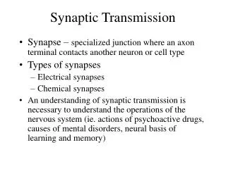
Synapse - Definition • From the Greek word synapsis which means junction. The anatomical relation of one nerve cell to another cell which allows impulse transmission (information exchange) between neurons, or between neurons and muscle or glands. Term coined by Sir Charles Sherrington.
Synapses Can Be Classified By: • Cytoarchitecture • Method of signal conduction (electrical/chemical) • Conductance of postsynaptic membrane to selective ion species (excitatory/inhibitory)
Classification of Synapses • Cytoarchitectural classification • Axo-dendritic synapse • Axo-somatic synapse • Axo-axonic synapse • Dendro-dendritic synapse • Soma-somatic synapse • Neuromuscular synapse (skel m.: NM junction) • Neuroglandular synapse
Classification of Synapses • Based on method of impulse conduction (electrical/chemical) • Electrical synapse (bridged or gap junction) - cytoplasmic continuity between pre- and postsynaptic elements allows direct conduction of electrical currents between cells by ion movements through connexons. Bidirectional. • Chemical synapse (unbridged junction) - thought to be the most numerous type of synapse. A chemical transmitter diffuses across a 20-30 nm synaptic cleft between cells. Unidirectional, with a delay of 0.3-5 msec.
Classification of Synapses • Based on method of impulse conduction (electrical/chemical) • Electrical synapse (bridged or gap junction) - cytoplasmic continuity between pre- and postsynaptic elements allows direct conduction of electrical currents between cells by ion movements through connexons. Bidirectional. • Chemical synapse (unbridged junction) - thought to be the most numerous type of synapse. A chemical transmitter diffuses across a 20-30 nm synaptic cleft between cells. Unidirectional, with a delay of 0.3-5 msec.
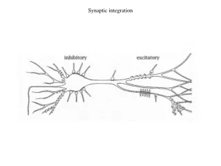
Classification of Synapses • Based on conductance of postsynaptic membrane to selective ion species (excitatory/inhibitory) • Excitatory synapse - an increase in postsynaptic membrane conductance to sodium, which depolarizes the membrane. • Inhibitory synapse - an increase in postsynaptic membrane conductance to potassium and/or chloride ions, which hyperpolarizes the membrane.
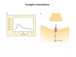
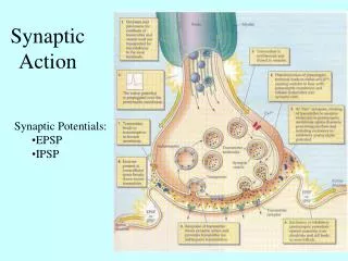
Excitatory post-synaptic potential • Action potential - Ca++ regulated release of neurotransmitter (NT) by synaptic vesicles • Attachment of NT to post-synaptic membrane receptors • Opening of ligand-gated channel to Na+ • Influx of Na+ makes the interior of the neuron more positive (excitatory post-synaptic potential, EPSP) • EPSP degrades with time and distance, moves toward neuron axon hillock
EPSP • Superimposed EPSP’s recorded from an a motor neuron (biceps-semitendinosus m.) in response to a repetitive shock to the afferent fibers of the Ia ending of its muscle spindle
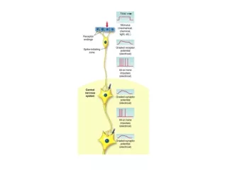
Initiation of an action potential by an EPSP • If an EPSP reaches the axon hillock or a spike initiation zone, and is sufficiently large to exceed threshold, an action potential is produced
Spatial Summation of EPSPs • Subliminal volleys from two afferents applied to the same target neuron, closely spaced in time
Temporal Summation of EPSP’s • Two well-spaced subliminal volleys to the same Ia fiber • Motor neuron response to same stimuli. Reduced interval EPSPs add up to produce an action potential
Spatial summation Temporal summation
EPSP Summation • Both temporal and spatial summation occur to produce a depolarization at the axon hillock which may or may not trigger an action potential
Classification of Synapses • Based on conductance of postsynaptic membrane to selective ion species (excitatory/inhibitory) • Excitatory synapse - an increase in postsynaptic membrane conductance to sodium, which depolarizes the membrane. • Inhibitory synapse - an increase in postsynaptic membrane conductance to potassium and/or chloride ions, which hyperpolarizes the membrane.
Inhibitory post-synaptic potential • Action potential - Ca++ regulated release of inhibitory neurotransmitter (NT) by synaptic vesicles • Attachment of NT to post-synaptic membrane receptors • Opening of ligand-gated channel to Cl-/K+ • K+ efflux/Cl- influx to make the interior of the neuron more negative (inhibitory post-synaptic potential, IPSP) • IPSP degrades with time and distance, moves toward neuron axon hillock
Inhibitory Post Synaptic Potential • IPSP recorded from an extensor motor neuron in response to stimulation of Ia fibers from the antagonistic flexor muscle
Temporal Summation of IPSPs • IPSPs sum when the interval between volleys is short • IPSPs reduce the membrane potential further below the critical firing level (threshold)
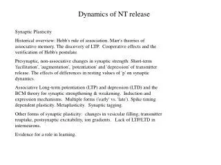
Origin/Distribution of Molecules Necessary for Synaptic Transmission • Molecules needed for synaptic transmission include enzymes, membrane constituents, and transmitter chemicals • Proteins are synthesized in the soma • Have to be transported down the axon • Metabolites must be transmitted back to the soma for elimination
Fast anterograde (orthograde) transport (400 mm/day) Vesicles, membranous organelles, certain neurotransmitters, enzymes, glycoproteins, precursors of receptors, lysosomes, mitochondria, growth factors Types of Axonal Transport
2. Axoplasmic flow (slow anterograde /orthograde) - 0.5-3 mm/day 3. Axoplasmic flow (slow retrograde)- 5-10 mm/day Biosynthetic and other soluble enzymes, neurofilament proteins, tubulins (for microtubules) breakdown products, actin, metabolic enzymes, viruses, tetnus toxoid, NGF, herpes, rabies Types of Axonal Transport
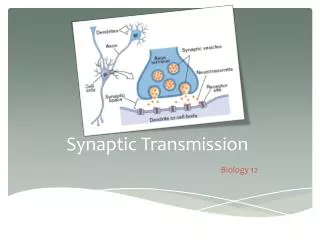
Steps in Synaptic Transmission • Depolarization of presynaptic membrane • Opening of voltage-gated Ca2+ channels • Ca2+ influx causes fusion of vesicles with presynaptic membrane • Neurotransmitter diffuses into synaptic cleft • Neurotransmitter binds to receptors on post-synaptic and presynaptic membranes • Neurotransmitter action terminated by (1) enzyme action in the cleft, (2) diffusion, or (3) re-uptake into presynaptic terminal
Typical Transmitter: ACh • First neurotransmitter identified, proving chemical neurotransmission • Otto Loewi (Austria, 1920’s) – studied innervation of the isolated frog heart by the vagus nerve • Experiment came to him in a dream – twice • Vagal stimulation slowed the heart; fluids bathing the first heart also slowed a second non-innervated heart • “Vagusstoff” – later renamed acetylcholine • Serendipity – seasonal variation in levels of AChE
Typical Transmitter: ACh cont. • ACh is synthesized in the cytoplasm of the presynaptic terminal • Precursors: choline and acetyl coenzyme A • Enzyme: choline acetyltransferase (ChAT) • Quaternary amine – charge limits crossing of membranes
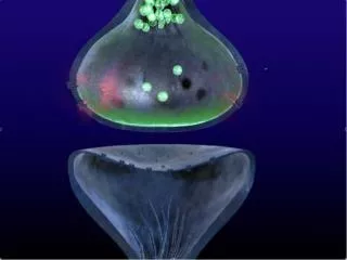
Typical Transmitter: ACh cont. • ACh is stored in synaptic vesicles in the presynaptic terminal • Some vesicles are free-floating, others are anchored against the presynaptic membrane • Ca2+ influx following depolarization causes vesicle fusion and NT release (in quanta) by exocytosis • Amount of NT released is proportional to amount of Ca2+ influx
Acetylcholine Metabolism acetylcholine ACh choline + acetate esterase (AChE) • AChE is located in the synaptic cleft • Choline is taken back up into the presynaptic terminal – active process • Acetate diffuses away to be utilized in • other metabolic roles
Typical Transmitter: ACh cont. • ACh is found in multiple locations: • Motor neurons to skeletal muscle (NMJ) • Autonomic nervous system neurons (PSNS, SNS) to smooth muscle and glandular tissue • Central nervous system – cortical arousal vs. sleep, Alzheimer’s disease, nicotine, etc.