Download
1 / 21
370 likes | 964 Views
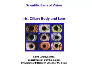
Iris, ciliary body and choroid. Iris. The iris lies in front of the lens and the ciliary body It separates the anterior chamber from the posterior chamber. Iris. The stroma connects a sphincter muscle (sphincter pupillae), which contracts the pupil

E N D
Iris • The iris lies in front of the lens and the ciliary body • It separates the anterior chamber from the posterior chamber
Iris • The stroma connects a sphincter muscle (sphincter • pupillae), which contracts the pupil • A set of dilator muscles (dilator pupillae) which open it. • Depending on the amount of light, the iris makes the pupil larger or smaller.
Structure of iris • The iris consists stroma. • The back surface is covered by an epithelial layer two cells thick (the iris pigment epithelium • The outer edge of the iris, known as the root, is attached to the sclera and the anterior ciliary body • . • The iris and ciliary body together are known as the anterior uvea
Aqueous humour Just in front of the root of the iris is the region through which the aqueous humour constantly drains out of the eye, with the result that diseases of the iris often have important effects on intraocular pressure, and indirectly on vision.
Blood supply and nerve supply • Constrictor muscles are innervated by parasympathetic fibers of the 3rd cranial nerve • Dilator muscles are innervated by sympathetic fibers from the superior cervical ganglion.
Disease of iris • Iritis Inflammation of the iris • Iris (uveitis) Busscca nodule Berlin nodule Koeppe nodule • Heterochromic iris • Iris atrophy • Aniridia • Iris cyst • Iris melanoma
Aniridia • It is usually bilateral condition in which the whole of the iris is appeared to be missing on external examination • The rudimentary iris, can be visible only by goniocopy • It is due failure of anterior growth and differentiation of the optic cup Symptoms • Photophobia • Nystagmus • Defective vision Treatment • Soft contact lens to reduce photophobia • Glaucoma responds poorly to treatment
Typical coloboma • The pupil is pear-shaped with broad base towards the pupillary margin. • Isolated typical coloboma of the iris is rare. It usually extends upto the ciliary body.
Albinism • This is a hereditary disorder of absence or reduction of the melanin pigmentation throughout the body. • The iris looks pink and translucent, owing to lack of pigment • The fundus appears orange pink in colour.
Heterochromic iris • It means the two irides show a significant difference in colour. • It may be congenital or acquired hepertic uveitis , trauma, acute attack of angle closure glaucoma.
Ciliary body • It lies between iris and choroid • It contains ciliary muscle and ciliary processes. • The ciliary muscle takes part in accommodation • Ciliary process produce aqueous humor. • Aqueous is transparent fluid which nourishes ocular structure
Flow of aqueous humor Ciliary process Posterior chamber Pupil Anterior chamber Trabecular meshwork Canal of schlemm Collector channel Episcleral veins
Blood and Nerve supply • Blood supply : by branches of long posterior ciliary artery and short anterior ciliary artery. • Nerve supply : Naso ciliary branch of 5th cranial nerve
Choroid • The vascular sheet separates the sclera from the retina. • It is 0.25mm of thick at posterior pole and 0.1mm thick anteriorly • There are 3 layer in the choroid • Outer vessel layer • middle vessel layer • Inner chorio-capillaries
Diseases of choroid • Choroiditis • Choroidal detachment • Choroidal coloboma
Choroiditis • Acute multifocal placoid pigment epitheliopathy (AMPPE) It is unilateral idiopathic disease affecting young adults. It is subacute loss of central vision with typical deep, cream colored placoid lesions • Investigations: FFA • Treatment : No effective treatment
Serpiginous choroiditis • It is a rare, idiopathic, recurrent disease of the RPE and chorio-capillaries disease. • Chorio retinal inflammatory lesion starts around the optic disc and spreads towards in all directions with out any inflammtory cells in the vitreous. • Investigations: FFA • Treatment : No effective treatment
Blood and nerve supply • Short posterior ciliary arteries • Two Long posterior ciliary arteries • Seven anterior ciliary arteries