Download
1 / 41
2.05k likes | 10.44k Views
Slit lamp biomicroscopy. OP1201 – Basic Clinical Techniques Part 1 Dr Kirsten Hamilton-Maxwell. Today’s goals. By the end of today’s lecture, you should be able to… List the uses of the slit lamp biomicroscope

E N D
Slit lamp biomicroscopy OP1201 – Basic Clinical Techniques Part 1 Dr Kirsten Hamilton-Maxwell
Today’s goals • By the end of today’s lecture, you should be able to… • List the uses of the slit lamp biomicroscope • Identify the main components of the slit lamp; be able to operate these components • Discuss and perform a series of basic illumination and magnification techniques • A second lecture will follow where we talk about combining these techniques into a routine and look at some examples
Why slit lamps are so great • Slit lamp assessment is considered to be the gold standard device for the assessment of the anterior segment of the eye in clinical practice • This is because they provide… • Excellent image quality • Stereoscopic image • Flexible illumination • Flexible magnification • Therefore there are many different uses • Even more when attachments are added
What can we use them for? On their own With accessories • Routine examination of anterior segment • Adnexa through to anterior vitreous • Problem-based examination of anterior segment • Contact lens examination • Assessment of anterior chamber depth and angle • Gonioscopy • Fundoscopy • Ocular photography • Contact tonometry (Goldmann) • Pachymetry • Corneal sensitivity measurements (aesthiometry) • Laser photocoagulation
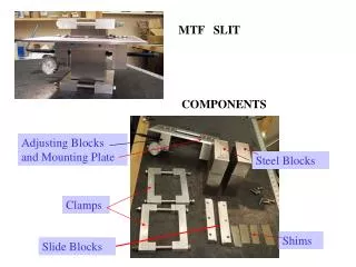
Basic Design • Viewing arm • Biomicroscope • Adjustable focus eyepieces • Magnification dial • Illumination arm • The “slit lamp” • Slit size, shape and filter controls • Variable size, shape, colour and brightness • Biomicroscope and illumination are mechanically coupled around central pivot point (copivotal) • Both focus at the same point (parfocal) • Both arms can swing independently 180º along horizontal – there is a scale in degrees • Both always central regardless of angle (isocentric) • Moveable base plate and joystick control
A good biomicroscope has… • Adequate working distance between the microscope and the eye to allow the practitioner to access the eye • Convenient size for use in practice • Adaptable to suit different practitioners • Good resolution • Good depth of focus • A wide range of magnifications
Magnification • Slit lamps provide variable magnification • Lower magnifications are used for general assessment and orientation • Higher magnifications are used for detailed inspections of areas of interest • There are several ways to do this • Common methods: Littmann-Galilean telescope and zoom systems • Less common methods: Change the eyepieces and/or change the objective lens
Littmann-Galilean telescope method • A separate optical system is placed in between the eyepiece and the objective • It consists of a rotating drum that house 2 Galilean telescopes plus a pair of empty slots • Optics refresher: Galilean telescopes consist of a positive and negative lens that provide magnification based on the lens powers and their separation • It is easy to identify whether the slit lamp you are using has this inside • The magnification dial will click into place as you turn it, and there will be numbers on the dial that correspond to the magnification in each position
A Galilean telescope Parallel light enters and exits. Magnification is typically the intended outcome. However, if you look from the other side, the image will be minified.
Two telescopes produce two magnifications • Mag highest when the convex lens is near objective • Reversal of these two telescopes produces two further minifications • No telescope provides 5th option
Zoom systems • This tends to be found on high-end Nikon, Topcon and Zeiss instruments • Magnification can vary between 7x to ~ 40X • I find that the image quality is not as good with zoom magnification
Change eyepieces or objective Eyepieces Objective • Often two sets provided with slit lamp • Typical values 10x, 12.5x, 15x or 20x • Inconvenient so rarely used • Generally unnecessary on modern slit lamps • Flip arrangement for rapid change • Usually only two options due to space confinements • Typical values are 1x and 2x Lever
Illumination The slit lamp
What makes a good slit? • A good slit needs to be • Bright • Evenly illuminated • Finely focused • Have well defined, straight edges • Flexible in terms of size, shape, colour and intensity • The illumination also needs to • Provide good colour rendering to detect subtle colour changes
Slit width • Continuously variable (0 to 12-14mm) • May be graduated to allow measurement • Narrow slits are used to “slice” through the cornea to determine depth or thickness • Wide slits are used to inspect surfaces
Slit height • May be continuous or set to fixed heights • Usually a combination of the two • May be graduated to allow for measurement • Long slits are used to view most structures in front of the pupil, while short slits pass through the pupil much better • Short slit also used to assess the clarity of the anterior chamber
Slit orientation • Achieved by rotating lamp housing
Filters • Slit lamps may have some/all of the following filters • Diffuser • Heat reduction • Neutral density • Polarising • Red free • Cobalt blue • Wratten (in observation system)
Methods of illumination • Direct • Indirect • Retro-illumination • Sclerotic scatter (next year) • Specular reflection (next year) • Conical section (next year) • A combination of these methods is used to view the anterior eye structures
Direct illumination The light and the microscope are both pointed at the object of interest Microscope Lamp
Direct illumination • There are several different forms, named simply by how wide the slit is • Diffuse (usually not a slit at all) • Wide beam • Parallelepiped • Optical section • The slit width will change what you can see • Diffuse/wide beam for an overall view • Wide parallelepiped for broad views of one plane (e.g. Surface of a structure) and narrow parallelepiped for a balanced view • Optical section to “cut through” a tissue, for thickness and depth
Effect of slit width (cornea) Wide beam: mostly surface Parallelepiped: balance of surface and depth Optical section: mostly depth
Why is the angle important • The angle between the microscope and the illumination arms is important. Wider angles… • Allow view of deeper layers without interference from reflections from upper layers • The wider the beam, the greater the angle needed to “see behind the surface layer” • Allows estimation of depth • Allows better perception of texture • Allows direct/indirect/retro simultaneously • You’ll find a graduated scale located at the pivot point of the two slit lamp arms • It will give you the total separation between the two arms in degrees
Effect of angle (cornea) 45º: balance of surface and depth 5º: surface only 85º: depth only
Wide beam/Diffuse • Used for general inspection of eye and adnexa • Good for colour assessment • Contact lens fit • Wide slit, diffusing inserted, microscope in front, illumination angle 30–50°, magnification of 6-10x • Patients are generally unable to tolerate the brightness of a wide beam This eye has iris naevi (freckles)
Parallelepiped • Default method for corneal inspection • Shows a block of tissue in 3-D, so good balance between surface and depth inspection • Beam about 2 mm, microscope/illumination, variable angle, medium to high mag (10-25x) This is a narrow parallelepiped being used to view iris and pupillary margin. The light first passes through the cornea but is out of focus there.
Optical section • Allows judgement of thickness or depth • Use the narrowest slit possible (0.1 – 0.2 mm), angled beam (largest angle possible), high illumination, and a dark room • You need very sharp focus A helpful tip: Even though this instrument is called a slit lamp, we hardly ever need to use a slit this narrow. Save it for when you need to work out the depth or thickness of a corneal lesion.
Indirect illumination An object being viewed is illuminated indirectly when it lit by reflections/scatter of light that occur when the light is shone other than onto the object itself. Microscope Lamp
Indirect illumination • Good for subtle detail, which would be obscured or washed out by large amounts of illumination • Light internally reflected within the cornea, or reflected by other surrounding tissue • Opacities scatter light so they will appear light in colour • They are best viewed against the dark pupil (or dark iris, if your patient happens to have one) • To achieve the effect, keep the slit width narrow to medium (2-4 mm), and view with a medium to wide angle. Magnification will vary depending on the size and extent of the object, but it’s typically medium to high for subtle defects
Directly illuminated Indirectly illuminated This picture shows a contact-lens related condition called neovascularisation. These are blood vessels in the cornea. In this example, we don’t move anything but our attention – the light and focus stays where it is. We can do this because the slit doesn’t light up our whole field of view
Retro-illumination An object of interest is lit by retro-illumination when the light source is directed onto another structure so that the reflected light must pass through that object. Microscope Lamp
Retro-illumination • Light may be reflected from 2 main structures: • Iris: this back-lights the cornea • Fundus: this back-lights the lens • Opacities will appear dark against a bright background • For iris retro-illumination, use a narrow-moderate width slit, a wide angle of illumination, and magnification appropriate to the object size/extent • Decoupling may be necessary when the magnification high • For fundus retro-illumination, use a short slit with narrow-moderate width, narrow angle of illumination (0-10º), and moderate magnification
Directly illuminated Retro-illuminated Indirectly illuminated This is the same example from earlier. The blood vessels in the indicated section are retro-illuminated because they are being lit from behind (the light has reflected off the iris).
This is an example of retro-illumination of the lens (the light has reflected from the retina). This patient has cortical spokes, which are indicative of early cortical cataract.
Marginal retro-illumination • At the border of the zones illuminated by indirect and retro, therefore viewing technique is similar for retro with high mag, decoupling helps • Objects of higher refractive index show “reversed illumination” • Useful to differentiate microcysts (high refractive index) from vacuoles (low refractive index)
Recommended reading • Elliott, Section 6.61-6.67. • There are lots of examples on Elliott Online (pictures and video) • For section 6.64, parts 2, 3, 4, 5 and 6 only