Download
1 / 20
200 likes | 374 Views
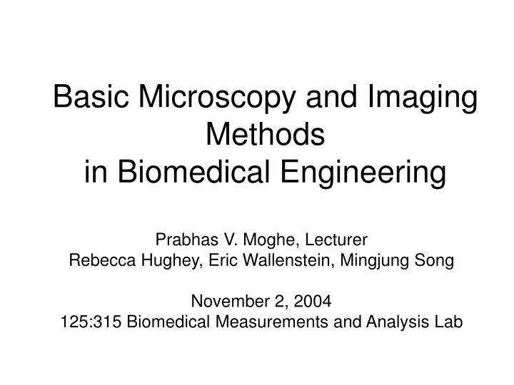
Basic Microscopy and Imaging Methods in Biomedical Engineering . Prabhas V. Moghe, Lecturer Rebecca Hughey, Eric Wallenstein, Mingjung Song November 2, 2004 125:315 Biomedical Measurements and Analysis Lab. General Resources

E N D
Basic Microscopy and Imaging Methods in Biomedical Engineering Prabhas V. Moghe, Lecturer Rebecca Hughey, Eric Wallenstein, Mingjung Song November 2, 2004 125:315 Biomedical Measurements and Analysis Lab
General Resources Primer in Microscopy by Olympus: http://www.olympusmicro.com/primer/opticalmicroscopy.html Molecular Expressions A comprehensive resource maintained by Florida State University. Includes a Microscopy Primer with tutorials on optical microscopy, digital imaging: http://micro.magnet.fsu.edu/primer/ microscopy.info An information source for microscopy and microanalysis including a hyperlinked glossary and links to professional societies and educational resources. http://www.mwrn.com World Wide Web Virtual Library: Microscopy: http://www.ou.edu/research/electron/www-vl/long.shtml Vast set of links covering all aspects of light, electron, and other types of microscopy, including extensive lists of imaging and microscopy labs, image galleries, and educational sites. Live Cell Imaging Applications in Confocal Microscopy: A lecture by Dr. J. Paul Robinson at Purdue Univ. http://www.cyto.purdue.edu/flowcyt/educate/confocal/524Lec11/sld001.htm NIH Office of Extramural Research: Links to Bioimaging Sites http://grants2.nih.gov/grants/bioimaging/links.htm Standard Microscopy Terminology: http://resolution.umn.edu/glossary/FrameGloss.html A comprehensive glossary of microscopy terms made available by the University of Minnesota Characterization Facility. Microscopy/Imaging Resources on WWW: http://swehsc.pharmacy.arizona.edu/exppath/micro/index.html A very well-organized, extensive collection of educational and informational resources, including links to numerous online image galleries, microscopy sites, and an introduction to digital imaging. * Science Magazine, April 2003, Special Issue on Biological Imaging WWW Bibliography -- Supplementary Review Materials*
Outline • Physics of Optical Microscopy Bright Field Microscopy Phase Contrast Microscopy Fluorescence Microscopy Confocal Microscopy • Biomedical Image Processing Image Acquisition Image Processing Steps Image Analysis • Cellular Properties Derived via Biomedical Imaging
Electromagnetic Spectrum The wavelength required to “see” an object must be the same size of smaller than the object
Physics of Optical Microscopy • The ability of a microscope objective to "grasp" the various rays coming from each illuminated part of the specimen is related to the angular aperture of the objective. N.A. = n . sin (u); n= refractive index; u=1/2 subtended angle - Max theoretical N.A. of a dry objective is 1 - Max theoretical N.A. of oil immersion objectives is 1.5
Optical Microscopy Issues: Resolution • Resolution is defined as the ability of an objective to separate clearly two points or details lying close together in the specimen. where R=resolution distance; l, the wavelength of light used; N.A. = the numerical aperture. - As N.A. increases, resolution gets better (R smaller). - Longer wave lengths yield poorer resolution.
Principle of Phase Contrast Microscopy • Zernicke: Greatest advance in Microscopy (1953) • Phase microscopy requires phase objectives and a phase condensor.
Fluorescence Microscopy Dichroic Mirror Barrier Filter Exciter Filter Mercury Light Source Objective/ Condensor Specimen
Microscopy to Image Cell Structure And Cell Architecture on Biomaterial Substrates Use of fluorescence microscopy and transmitted/reflected imaging Cytoskeletal Organization Rounded Phenotype Mildly Polarized Phenotype
Selected Basic Definitions in Biomedical Imaging & Image Analysis Pixels: 2-D Picture Elements Mean Gray Level: Gray-scale Intensity (0-256) MGL Histogram Segmentation and Thresholding (MGL Contrast) Filtering Densitometry Morphometry (Area, Shape factor, Polarity/Aspect Ratio)
Spatial and Gray Level Digital Resolution • Spatial resolution: • # of samples per unit length or area • DPI: dots per inch specifies the size of an individual pixel • If pixel size is kept constant, the size of an image will affect spatial resolution • Gray level resolution: • Number of bits per pixel • Usually 8 bits • Color image has 3 image planes to yield 8 x 3 = 24 bits/pixel • Too few levels may cause false contour
Example of Biomedical Image Processing • Biomedical polymer sponges were saturated with a fluorophore (fluorescein isothiocyanate - FITC) & imaged on a microscope using fluorescence microscopy. • Images of each porous field were digitized and stored on the computer. • Digitized images were analyzed using biomedical image processing to identify the number, shape, and location of pores. (Similar approaches can be used to identify fluorescently labeled cells and cell number, cell viability, cell morphology) Tjia and Moghe, J. Biomed. Mater. Res (Appl Biomat) 43: 291, 1998
Outline of the Lab ExercisesMicroscopy and Cellular Imaging • Learn how to : use a computer-interfaced optical microscope archive images process images and quantify results identify cells via fluorescence labeling of the cytoskeleton show cells are viable via fluorescence labeling of DNA visualize cell borders quantify cell morphology (phenotyping)