Download
1 / 1
10 likes | 59 Views
M19.24 consdabi@consdabi.org. Preliminary results on the genome instability evaluated by Micronucleus Test in four Italian pigs Ancient Autochthonous Genetic Types (AAGT) Matassino, D. 1,2 , Falasca, D. 1 , Gigante, G. 1 , Varricchio, G. 1 , Fornataro, D. 1

E N D
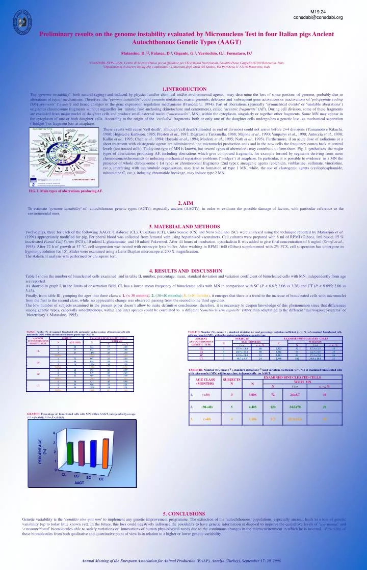
M19.24 consdabi@consdabi.org Preliminary results on the genome instability evaluated by Micronucleus Test in four Italian pigs Ancient Autochthonous Genetic Types (AAGT) Matassino, D.1,2, Falasca, D.1, Gigante, G.1, Varricchio, G.1, Fornataro, D.1 1ConSDABI. NFP.I.-FAO- Centro di Scienza Omica per la Qualità e per l'Eccellenza Nutrizionali, Località Piano Cappelle-82100 Benevento, Italy, 2Dipartimento di Scienze biologiche e ambientali - Università degli Studi del Sannio, Via Port'Arsa,11-82100 Benevento, Italy • INTRODUCTION • The ‘genome instability’, both natural (aging) and induced by physical and/or chemical and/or environmental agents, may determine the loss of some portions of genome, probably due to alterations of repair mechanisms. Therefore, the ‘genome instability’ could promote mutations, rearrangements, deletions and subsequent gene activations or inactivations of ‘polypeptide coding DNA segments’ (‘genes’) and hence changes in the gene expression regulation mechanisms (Franceschi, 1994). Part of aberrations (generally ‘symmetrical events’ or ‘unstable aberrations’) originates chromosome fragments without organelles for mitotic fuse anchoring (kinetochore and centromere), called ‘acentric fragments’ (AF). During cell division, some of these fragments are excluded from major nuclei of daughter cells and produce small external nuclei (‘micronuclei’, MN), within the cytoplasm, singularly or together other fragments. Some MN may appear in the cytoplasm of one or both daughter cells. According to the origin of the ‘excluded’ fragments, both or only one of the daughter cells undergo/oes a genetic loss: as mechanical separation (‘bridges’) or fragment loss at anaphase. These events will cause ‘cell death’, although‘cell death’(intended as end of division) could not arrive before 2÷4 divisions (Yamamoto e Kikuchi, 1980; Högsted e Karlsson, 1985; Preston et al., 1987; Degrassi e Tanzanella, 1988; Majone et al., 1990; Vanparys et al., 1990; Antoccia et al., 1990; Kallio et al., 1993; Chen et al., 1994; Hayashi et al., 1994; Modesti et al., 1995; Nath et al., 1995). Furthermore, if an acute dose of radiations or a short treatment with clastogenic agents are administered, the micronuclei production ends and in the new cells the frequency comes back at control levels (not treated cells). Today one type of MN is known, but several types of aberrations may contribute to form them. Fig. 1 synthetizes the major types of aberrations producing AF,including aberrations which give compound fragments, for example formed by segments deriving from more chromosomes/chromatids or inducing mechanical separation problems (‘bridges’) at anaphase.In particular, it is possible to evidence in a MN the presence of whole chromosome ( 1st type) or chromosomal fragments (2nd type); aneugenic agents (colchicin, vinblastine, sulfunate, vincristine, ecc.), interfering with microtubule organization, may lead to formation of type 1 MN, while, the use of clastogenic agents (cyclophosphamide, mitomicine C, ecc.), inducing chromatide breakage, may induce type 2 MN. FIG. I. Main types of aberrations producing AF. 2. AIM To estimate ‘genome instability’ of autochthonous genetic types (AGTs), especially ancient (AAGTs), in order to evaluate the possible damage of factors, with particular reference to the environmental ones. 3. MATERIAL AND METHODS Twelve pigs, three for each of the following AAGT: Calabrese (CL), Casertana (CT), Cinta Senese (CS) and Nero Siciliano (SC) were analysed using the technique reported by Matassino et al. (1994) appropriately modified for pig. Peripheral blood was collected from femoral vein using heparinized vacutainers. Cell cultures were prepared with 8 ml of RPMI (Gibco), 1ml blood, 15 % inactivated Foetal Calf Serum (FCS), 10 ml/ml L-glutammine and 10 ml/ml Pokeweed. After 44 hours of incubation, cytochalisin B was added to give final concentration of 6 mg/ml (Scarfì et al., 1993). After 72 h of growth at 37 °C, cell suspension was treated with eritrocyte lysis buffer. After washing in RPMI 1640 (Gibco) supplemented with 2% FCS, cell suspension has undergone to hypotonic solution for 15’. Slides were examined using a Leitz Diaplan microscope at 200 X magnification. The statistical analysis was performed by chi square test. 4. RESULTS AND DISCUSSION Table I shows the number of binucleated cells examined and in table II, number, percentage, mean, standard deviation and variation coefficient of binucleated cells with MN, independently from age are reported. As showed in graph I, in the limits of observation field, CL has a lower mean frequency of binucleated cells with MN in comparison with SC (P < 0.01; 2.06 vs 3.26) and CT (P < 0.005; 2.06 vs 3.43). Finally, from table III, grouping the ages into three classes:1. (< 30 months),2. (30÷40 months);3. (>40 months),it emerges that there is a trend to the increase of binucleated cells with micronuclei from the first to the second class, while no appreciable change was observed passing from the second to the third age class. The low number of subjects examined in the present paper doesn’t allow to make definitive conclusions; therefore, it is necessary to deepen knowledge of this phenomenon since that differences among genetic types, especially autochthonous, within and inter species could be correlated to a different ‘constructivism capacity’ rather than adaptation to the different ‘microagroecosystems’ or ‘bioterritory’ ( Matassino, 1995). GRAPH I. Percentage of binucleated cells with MN within AAGT, independently on age (** = P<0.01; ***= P < 0.005). 5. CONCLUSIONS Genetic variability is the ‘conditio sine qua non’ to implement any genetic improvement programme. The extinction of the ‘autochthonous’ populations, especially ancient, leads to a loss of genetic variability (up to today little known yet). In the future, this loss could negatively influence the possibility to have genetic information at disposal to improve the qualitative levels of ‘nutritional’ and ‘extranutritional’ biomolecules able to satisfy variations or innovations of human physiological needs due to the continuous changes in the microenvironment in which he is inserted. Variability of these biomolecules from both qualitative and quantitative point of view is in relation to a higher or lower genetic variability. Annual Meeting of the European Association for Animal Production (EAAP), Antalya (Turkey), September 17÷20, 2006