Download
1 / 5
50 likes | 57 Views
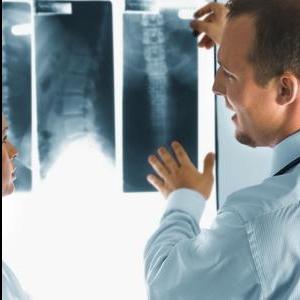
There are several different types of equipment and radiological methods for brain imaging. These techniques can be used to detect diseases such as Parkinsonu2019s disease and Alzheimeru2019s disease. They can also be used to detect lesions without having to perform surgery. Wessam Bou-Assaly studied neuroradiology at the Indiana University, School of Medicine. He is a skilled radiologist who has years of experience in neuroradiology as well as nuclear medicine. <br>For more updates and Wessam Bou-Assaly click here.. https://wessambouassaly0.wordpress.com/<br>

E N D
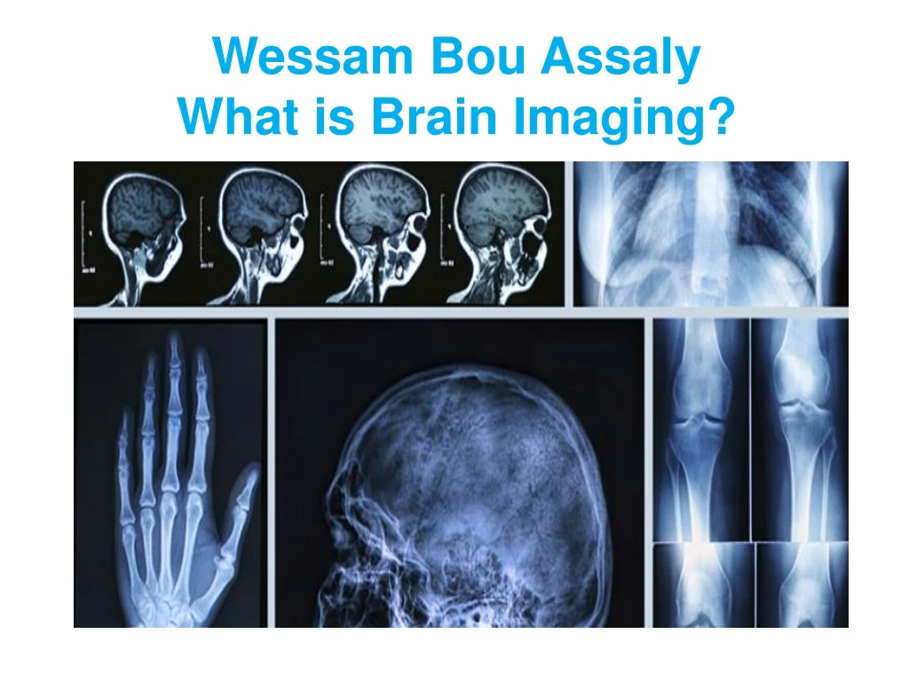
Wessam BouAssaly What is Brain Imaging?
Wessam Bou-Assaly Brain imaging, or neuroimaging, is the process of creating an image of the brain that can be used to study the structure and function of the organ. Wessam Bou-Assalyis a radiologist who specializes in neuroradiology and nuclear medicine.
Wessam Bou-Assaly He spent seven years working as an Assistant Professor of Radiology in the neuroradiology division of the University of Michigan, Department of Radiology. He is experienced in neuroimaging.
Wessam Bou-Assaly What is Brain Wessam BouAssaly:- There are several methods used to create images of the brain. Computed tomography (CT) scanning is a common way to create an image of the brain. This process includes placing an x-ray source on a large ring and having the patient lay with their head inside that structure. X-rays are then projected over the patient’s head and an image is created based on the absorption of those rays. Another way to create a brain image is to use Positron Emission Tomography (PET). This method involves nuclear medicine. The patient either ingests small amounts of radioactive substances, or the substances are ingested.
THANK YOU... For more details visit at : http://wessambouassaly.blogspot.com/ https://www.linkedin.com/in/wessam-bou-assaly-89428526 https://www.pinterest.com/wessambouassaly/ http://www.slideshare.net/wessembouassaly