Download
1 / 24
240 likes | 396 Views
INTRODUCTION TO ARTHOROLGY. Kaan Yücel M.D., Ph.D . 6.January.2 01 4 Monday. 1.1. CLASSIFICATION OF JOINTS 1.2. STABILITY OF JOINTS 1.3. JOINT VASCULATURE AND INNVERVATION. Arthrology Greek a rqron joint – logy. science concerned with the

E N D
INTRODUCTION TO ARTHOROLGY Kaan Yücel M.D., Ph.D. 6.January.2014 Monday
1.1. CLASSIFICATION OF JOINTS 1.2. STABILITY OF JOINTS 1.3. JOINT VASCULATURE AND INNVERVATION
Arthrology Greek a rqron joint –logy • science concerned with the • anatomy, function, dysfunction and treatment of joints.
according to the tissues that lie between the bones: Fibrous joints Cartilaginous joints Synovial joints Classification of Joints
Fibrous joints • Bones are united by fibrous tissue. • Suturesof the cranium
Fibrous joints • Syndesmosistype of fibrous joint • unites the bones with a sheet of fibrous tissue • either a ligament or a fibrous membrane • partially movable • The interosseous membrane in the forearm is a sheet of fibrous tissue that joins the radius and ulna in a syndesmosis.
Fibrous joints Dentoalveolarsyndesmosis (gomphoses or socket) a peglike process fits into a socket articulation between the root of the tooth and the alveolar process of the jaw.
Cartilaginous joints Bones are united by hyaline cartilage or fibrocartilage.
Cartilaginous joints Pimarycartilaginous joints-synchondroses hyaline cartilage- growth of a bone duringearly life Secondary cartilaginous joints-symphyses strong, slightly movable joints united by fibrocartilage
Synovial joints • Most common type of joints • Bones united by a joint capsule enclosing an articular cavity. • Providefree movement between the bones they join. • Joint cavity • potential space • contains lubricating synovial fluid, secreted by the synovial membrane. • Articular cartilage • articular surfaces are covered by hyaline cartilage • Articular capsule • surrounds the joint and formed of two layers.
Joint (articular) capsule • surrounds the joint • two layers. • Fibrous membrane • Synovial membrane
Synovial membrane • lines inner surface of the fibrous membrane. • highly vascular • produces synovial fluid, and lubricates the articulating surfaces • (helps to minimize the friction by articular surfaces). • attaches to the margins of the joint surfaces at the interface between the cartilage and bone and encloses the articular cavity.
Fibrous membrane Formedby dense connective tissue Surroundsand stabilizes the joint. Parts of it may thicken to form ligaments, further stabilize the joint. Ligaments outside the capsule usually provide additional reinforcement.
Closed sacs of synovial membrane also occur outside joints where they form synovial bursaeor tendon sheaths.
Ligaments • a cordorband of connectivetissueunitingtwostructures. • Articularcapsulesareusuallystrengthenedbyarticularligaments. • Connect thearticulatingbonestoeachother. • limit theundesiredand/orexcessivemovements of thejoints.
Articulardisc: Help toholdthebonestogether. Labrum: A fibrocartilaginous ring whichdeepensthearticularsurfaceforone of thebones.
Bursa • Flattenedsacsthatcontainsynovialfluidtoreducefriction. • Wallsare separated by a film of viscous fluid. • Foundwherever tendons rub against bones, ligaments, or other tendons.
Stability of Joints depends on four main factors negative pressure within the joint cavity shape, size, and arrangement of the articular surfaces ligaments tone of the muscles around the joint
Jointvasculatureandinnvervation • Joints receive blood from articular arteries that arise from the vessels around the joint. • Articular veins are communicating veins that accompany arteries (L. venae comitantes) and, like the arteries, are located in the joint capsule, mostly in the synovial membrane. • Joints have a rich nerve supply provided by articular nerves with sensory nerve endings in the joint capsule.
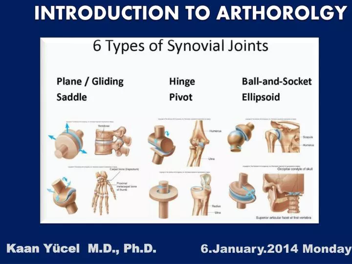
Types of synovial joints • according to shape of articulating surfaces- type of movement they permit • Plane joints • uniaxialjoints- glidingorsliding • acromioclavicular joint • 2. Hinge joints • uniaxialjoints- flexion & extension • knee & elbowjoints
Types of synovial joints 3. Saddle joints biaxialjoints- flexion & extension, abduction & adduction carpometacarpal joint at the base of the 1st digit (thumb) 4. Condyloid (ellipsoid type) biaxialjoints- flexion & extension, abduction & adduction metacarpophalangeal joints (knuckle joints) radiocarpal joint (wrist)
Types of synovial joints 5. Ball and socket joints (spheroidaljoints) multiple axes and planes: flexion and extension, abduction and adduction, medial and lateral rotation, and circumduction hip & shoulderjoints
Types of synovial joints 6. Pivot joints uniaxialjoints- rotationaround a centralaxis proximal& distal radioulnar joints